Part 4: Atherosclerosis
Mon Jul 20 2026
By B. Hassan
Atherosclerosis, commonly known as “hardening of the arteries”, is the leading cause of morbidity and mortality in developed nations. Through its major manifestations of myocardial infarction and stroke, it has become a major cause of death.
Normal Arterial Wall
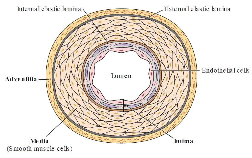
The arterial wall consists of three layers:
-
The intima, closest to the arterial lumen and therefore most “intimate” with the blood
-
The middle layer, known as the media
-
The outer layer, also called the adventitia
A single layer of endothelial cells covers the inner intimal surface and provides a barrier between circulating blood and the vessel wall.
The media is the thickest layer of the normal artery. It consists of smooth muscle cells and extracellular matrix. It is separated from the intima by an elastic boundary known as the internal elastic lamina and separated from the adventitia by an elastic boundary known as the external elastic lamina. The smooth muscle cells serve the contractile function of the artery, controlling resistance of the vessels to blood flow. The elastic layers function to stretch during the high pressure of systole and then recoils during diastole, propelling blood forward and serving a dampening function; keeping the blood flowing while the heart is in diastole (i.e. not actively pushing blood).
Generally, the elastic component is more prominent in larger arteries like the aorta and its direct branches, while the muscular component is more prominent in smaller arteries and arterioles.
Note
Due to their high smooth muscle content, arterioles provide approximately 80% of the total resistance to blood flow through the entire human circulation. This acts as a “dam” system, shielding the fragile single-cell-thick capillary beds right behind them from high pressure so they don’t rupture.
The adventitia contains nerves, lymphatics, and small blood vessels (vasa vasorum) that nourish the cells of the arterial wall. These are more prominent in larger arteries and disappear gradually as the vessels get smaller.
Endothelial cells
In a healthy artery, the endothelium performs structural, metabolic, and signaling functions that maintain homeostasis of the vessel wall.
The tightly adjoined endothelial cells form a barrier that contains blood within the lumen of the vessel and controls the passage of large molecules from the circulation into the subendothelial space.
As blood traverses the vessels, it encounters anti-thrombotic molecules produced normally by the endothelium that prevent it from clotting or that promote fibrinolysis (the breakdown of fibrin clots). Some of these molecules reside on the endothelial surface, including heparan sulfate, thrombomodulin, and tissue plasminogen activators. Others are secreted into the circulation by the endothelium to oppose other circulating pro-thrombotic substances; these include prostacyclins and nitric oxide (NO).
Although a net anti-coagulant state normally prevails, the endothelium can also produce pro-thrombotic and anti-fibrinolytic molecules when subjected to various stressors like a local injury.
Furthermore, endothelial cells secrete substances that modulate contraction of the underlying smooth muscles. These substances include vasodilators (e.g. NO and prostacyclin) and vasoconstrictors (e.g. endothelin) that alter the arteriolar resistance and therefore blood flow. In a normal artery, the predominance of vasodilator substances results in net smooth muscle relaxation.
Endothelial cells can also modulate the immune response. In the absence of pathologic stimulation, endothelial cells resist leukocyte adhesion and thus produce a local anti-inflammatory effect. However, endothelial cells respond to local injury or infection by expressing cell surface adhesion molecules, which attach immune cells to the endothelium, and chemokines, which attract leukocytes to the site of injury.
Smooth muscle cells
Smooth muscle cells synthesize the collagen, elastin, and proteoglycans that form the bulk of the vascular extracellular matrix. In addition, they produce vasoactive and inflammatory mediators, including interleukin-6 (IL-6) and tumor necrosis factor (TNF).
In normal arteries, most smooth muscle cells reside in the medial layer, with some smooth muscle cells in the intima, particularly in sites predisposed to atherosclerosis. During atherogenesis, medial smooth muscle cells can migrate into the intima, proliferate, and augment synthesis of extracellular matrix.
Extracellular Matrix
Collagen, elastin, and proteoglycans make up most of the extracellular matrix, which mostly resides in the medial layer. Interstitial collagen fibrils possess great tensile strength, while elastin provides flexibility. Together these components maintain the structural integrity of the vessel to withstand the high pressure within the lumen.
The extracellular matrix also regulates the growth of its resident cells. Collagen, in particular, can inhibit smooth muscle cell proliferation in vitro.
Lipoproteins
Lipoproteins are specialized transport particles that carry water-insoluble (hydrophobic) substances like lipids and cholesterol through the bloodstream. They consist of a central lipid core containing water-insoluble fats and a more hydrophilic (water-loving) outer layer composed of phospholipids, free cholesterol, and apolipoproteins (also called apoproteins). The apoproteins present on various classes of lipoprotein molecules act as the system’s conductors, directing the lipoproteins to specific tissues and mediate essential enzymatic reactions.
Lipoproteins are classified into five major groups based on their densities, lipid constituents, and associated apoproteins. In order of increasing density (and decreasing size), they are:
-
Chylomicrons (Lowest density, largest size)
-
Very-Low-Density Lipoproteins (VLDL)
-
Intermediate-Density Lipoproteins (IDL)
-
Low-Density Lipoproteins (LDL)
-
High-Density Lipoproteins (HDL) (Highest density, smallest size)
Exogenous (Intestinal) Pathway
This pathway processes fats that enter the body from external dietary sources.
-
Dietary fats are absorbed by the small intestine and repackaged into chylomicrons, accompanied by apo B-48. These are large particles particularly rich in triglycerides.
-
Once in the bloodstream, HDL donates apo E and subtypes of apo C to the chylomicrons.
-
Apo C (specifically the CII subtype) activates the enzyme lipoprotein lipase, which is located on the endothelial surface of adipose and muscle tissues. This enzyme hydrolyzes the chylomicrons, releasing free fatty acids. Adipose tissue uses these free fatty acids for storage, while cardiac and skeletal muscles use them for energy.
-
The stripped-down chylomicron remnants are recognized via apo E and removed from circulation by the liver.
-
Inside the liver, one fate of this cleared cholesterol is its incorporation into bile acids. Another fate is the export and usage of the cholesterol for synthesis of steroid hormones.
The endogenous pathway
Because dietary fat availability is not constant, the endogenous pathway provides a reliable supply of triglycerides for tissue energy needs.
The liver packages cholesterol and triglycerides into VLDL particles, accompanied by apo B-100. While VLDL’s triglyceride content is much higher than its cholesterol content, this is the main method the liver uses to release cholesterol into circulation.
Lipoprotein lipase breaks down VLDL, releasing fatty acids to muscle and adipose tissues. During this process, VLDL interacts with HDL, exchanging some of its triglycerides for apo C subtypes and apo E.
The breakdown of VLDL leaves behind intermediate-density lipoproteins (IDL), which faces two fates:
-
Approximately 50% of IDL remnants are directly cleared in the liver by hepatic receptors that recognize apo E.
-
The remaining IDL is catabolized further by lipoprotein lipase and hepatic lipase. These enzymes remove apo E and apo C, transforming the IDL into LDL particles.
Plasma clearance of the remaining LDL occurs primarily via LDL receptor-mediated endocytosis in both the liver and peripheral cells, a process directed by LDL’s apo B-100 and apo E.
Cellular Cholesterol Homeostasis
Intracellular cholesterol content is tightly balanced via a feedback loop involving de novo synthesis, cellular uptake, storage, and cellular efflux.
When internal cellular cholesterol levels drop, the cell triggers a mechanism to make and capture more cholesterol; the transcription factor SREBP (sterol regulatory element–binding protein) is released from the endoplasmic reticulum and enters the nucleus to increase the transcription of:
-
HMG-CoA Reductase: The absolute rate-limiting enzyme of cholesterol biosynthesis.
-
The LDL Receptor: The primary controller of cellular cholesterol uptake from the bloodstream via receptor-mediated endocytosis of circulating LDL.
Intracellular cholesterol excess
When a cell experiences an overabundance of internal cholesterol, it triggers a defense mechanism to push the excess out; the cell increases the transcription of the ABCA1 and ABCG1 genes (ATP-binding cassette A1 and G1).
The ABCA1 gene codes for a transmembrane protein transporter that pumps cholesterol out of the cell, forming nascent (immature) HDL particles. The product of the ABCG1 gene facilitates further efflux of cholesterol through this transmembrane protein transporter.
As circulating HDL picks up this free cholesterol, the cholesterol is esterified by the enzyme LCAT (lecithin cholesterol acyltransferase), which is activated by apo AI.
Note
The esterification of HDL is the process by which free cholesterol on the surface of HDL is converted into cholesteryl esters, increasing the cholesterol’s hydrophobicity, forcing it to move into the core of the HDL particle. This transition causes the HDL to change from a flat disc shape to a mature, spherical particle.
To get that excess cholesterol back to the liver, the body uses two routes:
-
Most cholesteryl esters in HDL are exchanged for triglycerides with any of the apo B-containing lipoproteins (VLDL, IDL, LDL), which then deliver it back to the liver.
-
HDL can also deliver cholesterol directly to the liver and steroid-producing tissues via the SR-B1 scavenger receptor.
ApoA-I is the structural backbone of HDL. Once HDL has been stripped of its lipid core via the routes described above, its protein components face two different clearance mechanisms:
-
As HDL drops off its fat cargo, it shrinks back down into a very small “lipid-poor” particle. The ApoA-I proteins easily detach from the shrinking particle where they are filtered out by the kidneys.
-
Instead of just emptying the fat cargo, the liver can also uptake the entire mature HDL particle whole via specific endocytic receptors. Once inside the liver cell, the entire unit is completely broken down in the cell’s lysosomes.
Atherosclerotic Arterial Wall
With the recognition that vascular wall cells respond to, and produce, pro-inflammatory agents, investigations into the role of endothelial and smooth muscle cells in atherogenesis have been ongoing. This research has identified several key components that contribute to the atherosclerotic process, including endothelial dysfunction, accumulation of lipids within the intima, recruitment of leukocytes, smooth muscle cell proliferation, formation of foam cells, and deposition of extracellular matrix.
Rather than follow a clear sequential path, the cells of atherosclerotic lesions continuously interact and modify each other’s behavior, shaping the plaque over decades into one of many possible profiles. These mechanisms can be categorized into 3 pathologic stages: the fatty streak, plaque progression, and plaque disruption. Lesions of all 3 stages can coexist, often side by side.
Fatty streak formation
Fatty streaks represent the earliest visible lesions in atherosclerosis. Although the precise inciting event of fatty streak development is not known, observations in animals suggest that various stressors cause early endothelial dysfunction. Such dysfunction initiates a series of distinct pathological stages culminating in the formation of advanced, complex fibro-fatty plaques.
Endothelial Dysfunction
Arterial endothelial injury and subsequent dysfunction serve as the primary inciting events that initiate atherosclerosis. This dysfunction can result from a combination of physical forces and chemical irritants.
Straight sections of arteries experience normal laminar flow and high shear stress. This activates transcription factors like Krüppel-like factor 2 (KLF2), which serves an “atheroprotective” function by accentuating the expression of the antioxidant enzymes like superoxide dismutase. Laminar forces also favor endothelial production of NO, an endogenous vasodilator, inhibitor of platelet aggregation, and anti-inflammatory substance. Consequently, arteries with few branches (such as the internal mammary artery) show a relative resistance to plaque development.
Arterial branch points and bifurcations, on the other hand, experience disturbed blood flow and low shear stress, which impairs these locally atheroprotective endothelial functions. Accordingly, bifurcated vessels (such as the common carotid and left coronary arteries) are common sites for plaque formation.
Endothelial dysfunction may also result from exposure to chemical stressors. Tobacco smoking, abnormal circulating lipid levels, and diabetes all increase the endothelial production of reactive oxygen species (ROS), which directly promotes local vascular inflammation.
Lipoprotein Entry and Modification
Once the endothelium is dysfunctional, it no longer serves as an effective barrier to circulating elements, triggering a cascade of accumulation and chemical alteration.
Infiltration: Increased endothelial permeability allows LDL to easily enter the intima. This process is heavily accelerated by elevated circulating LDL concentrations seen in patients with hypercholesterolemia.
Note
High circulating LDL stems partly from diet, but it can also be driven by specific genetic mutations affecting the LDL receptor, apolipoprotein B, or PCSK9 (a protease involved in regulating the LDL receptor).
Retention: Once inside the intima, LDL accumulates in the subendothelial space by binding to proteoglycans within the extracellular matrix. This trapping increases the residence time of LDL inside the vessel wall. Hypertension, a major cardiovascular risk factor, further promotes this retention by stimulating smooth muscle cells to produce even more of these LDL-binding proteoglycans.
Chemical Modification: Once trapped within the subendothelial space, the sequestered LDL is physically cut off from plasma antioxidants and undergoes modifications:
-
Oxidation: Driven by local reactive oxygen species, prooxidant enzymes, or infiltrating macrophages, transforming the particles into oxidized LDL.
-
Glycation: In diabetic patients, excess glucose leads to the glycation of LDL, another distinct modification that ultimately renders the lipoprotein highly pro-inflammatory.
Leukocyte Recruitment
The presence of modified LDL and local inflammation signals the immune system, initiating the recruitment of leukocytes (primarily monocytes and T lymphocytes) into the vessel wall. This process depends on the expression of leukocyte adhesion molecules and on chemoattractant signals that direct leukocytes into the subintimal space.
Two major subsets of leukocyte adhesion molecules persist in the inflamed atherosclerotic plaque:
-
The immunoglobulin gene super family, especially vascular cell adhesion molecule-1 (VCAM-1) and intercellular adhesion molecule-1 (ICAM-1)
-
The selectins, particularly E-selectin and P-selectin.
Chemoattractant signals, such as monocyte chemotactic protein-1 (MCP-1), create a chemical gradient that directs diapedesis (the passage of white blood cells through the intact endothelial layer) into the subintimal space.
Modified LDL and pro-inflammatory cytokines can induce the expression of these adhesion molecules and chemoattractants independently. However, modified LDL also stimulates neighboring endothelial and smooth muscle cells to produce more pro-inflammatory cytokines, reinforcing the direct action. This dual ability to promote leukocyte recruitment and inflammation both directly and indirectly persists throughout the entire atherogenic process.
Foam Cell Formation
Once monocytes cross into the subintimal space, they transform into macrophages and begin consuming the modified lipids, giving rise to the earliest visible stages of the disease.
Classic LDL receptors do not recognize modified LDL particles. Instead, macrophages rely on a specialized family of scavenger receptors that preferentially bind modified LDL. Unlike the classic receptor, uptake via scavenger receptors completely evades negative feedback inhibition; the cells will continue to ingest lipids without stopping.
While this massive uptake may provide initial benefits by temporarily sequestering potentially damaging modified LDL particles, the rate of lipid influx vastly outpaces the cells’ ability to clear them (impaired efflux). As these macrophages become engorged with cholesteryl esters, they transform into foam cells. These foam cells serve as a continuous source of additional pro-inflammatory chemicals, further increasing local monocyte recruitment.
The localized grouping of these lipid-laden foam cells creates a fatty streak, representing the earliest visible lesion in atherosclerosis. On gross inspection, they appear as yellow areas of discoloration on the artery’s inner surface. They do not cause symptoms, do not protrude substantially into the lumen, and do not impede blood flow.
Note
Surprisingly, fatty streaks exist in the arteries of most people by age 20. They do not cause symptoms, and in some specific locations, they may regress over time.
Plaque progression
Whereas endothelial cells play a central role in the formation of the fatty streak, smooth muscle cells in the intima promote plaque progression by producing extracellular matrix that traps lipoproteins and adds to the bulk of the lesion. Not all fatty streaks progress into advanced lesions, and it is unknown why some evolve and others do not.
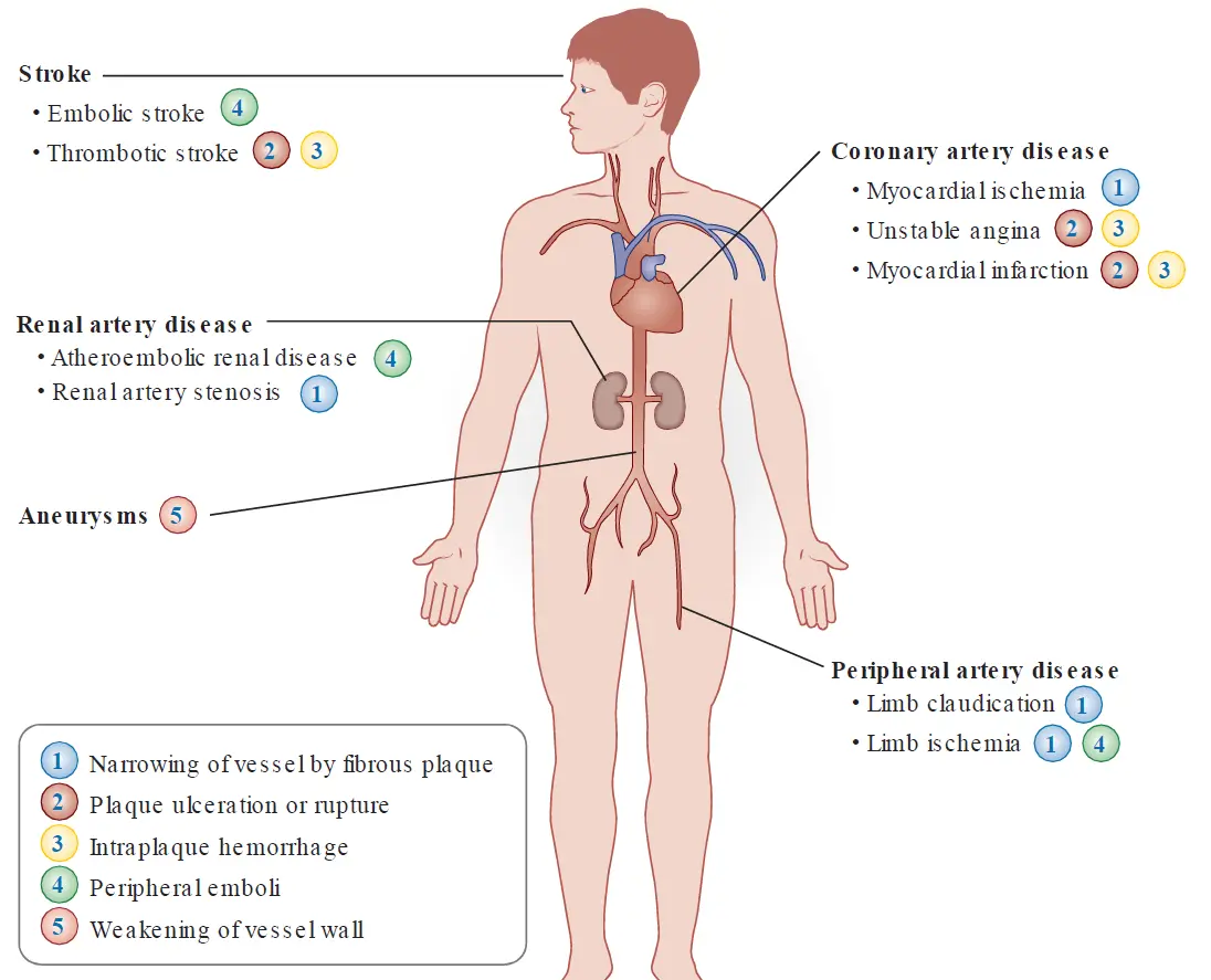
Early plaque growth typically involves a compensatory outward remodeling of the arterial wall that preserves the diameter of the lumen and permits plaque accumulation without limiting blood flow (no ischemic symptoms). Lesions at this stage can thus evade detection by angiography. Later plaque growth, however, can outpace the compensatory arterial enlargement, restrict the vessel lumen, and impede perfusion. Such plaques can result in tissue ischemia, causing symptoms such as angina pectoris or intermittent claudication of the extremities.
Many acute coronary syndromes (acute myocardial infarction and unstable angina pectoris) result when the fibrous cap of an atherosclerotic plaque ruptures, exposing prothrombotic molecules within the lipid core and precipitating an acute thrombus that suddenly occludes the arterial lumen.
One form of advanced plaques happens over time when the clearance of dead foam cells becomes inefficient. This leads to a breakdown of these cells, promoting the chronic accumulation of cellular debris and free extracellular lipids in the deep layer of the intima, forming what is called a necrotic core.
Another form of advanced lesions is known as fibro-fatty lesion.
Fibro-fatty lesion
The transition from fatty streak to fibrous plaque involves the migration of smooth muscle cells from the arterial media into the intima, where they then proliferate and secrete extracellular matrix molecules.
Foam cells, endothelial cells, and platelets entering through micro-fissures in the plaque surface can all elaborate substances that signal smooth muscle migration and proliferation. For example, they release platelet-derived growth factor (PDGF), which likely stimulates the migration of smooth muscle cells across the internal elastic lamina and into the subintimal space, where they subsequently replicate. PDGF additionally stimulates the growth of resident smooth muscle cells in the intima. Foam cells also release cytokines and growth factors (e.g., TNF, IL-1, fibroblast growth factor, and TGF-β) that further incite smooth muscle cell proliferation and/or the synthesis of extracellular matrix proteins. Moreover, these cytokines induce smooth muscle and leukocyte activation, promoting further cytokine release, thus reinforcing and maintaining the inflammation cycle.
This elaboration of extracellular matrix causes structural shifts that drive the evolution of the simple fatty streak into a complex fibro-fatty lesion.
Traditionally, plaques were thought to grow gradually and continuously, but current evidence suggests that this progression may be punctuated by subclinical events with bursts of smooth muscle replication. For example, morphologic evidence of intraplaque hemorrhages indicates that breaches in plaque integrity can occur without clinical symptoms or signs. Such plaque disruptions exposes tissue factor from foam cells to blood, which activates coagulation resulting in a microthrombus formation. Activated platelets within such microthrombi release additional factors (including PDGF) that can spur a local wave of smooth muscle migration and proliferation.
Activated T cells also contribute to plaque evolution. Th1 cells produce proinflammatory cytokines that promote plaque progression and instability, while Th2 lymphocytes and regulatory T cells produce factors, including TGF-β and IL-10, which can inhibit smooth muscle proliferation and potentially mitigate plaque growth.
As the predominant collagen-synthesizing cell type, smooth muscles favor fortification of the fibrous cap. Net matrix deposition depends on the balance of its synthesis by smooth and its degradation, mediated in part by a class of proteolytic enzymes known as matrix metalloproteinases. While PDGF and TGF-β stimulate production of collagens by smooth muscles, the Th1-derived cytokine interferon-γ (IFN-γ) inhibits smooth muscle collagen synthesis. Furthermore, inflammatory cytokines stimulate foam cells to secrete matrix metalloproteases, thereby weakening the fibrous cap and predisposing it to rupture.
Plaque disruption
The tug-of-war between matrix synthesis and degradation continues over decades but not without consequences. Death of smooth muscle and foam cells, either owing to excess inflammatory stimulation or by activation of apoptosis pathways, liberates cellular content, adding lipids and cellular debris to the growing lipid core. As the size of the lipid core increases, it protrudes into the arterial lumen, and mechanical stress focuses on the plaque border called the shoulder region.
The structure of the fibrous cap contributes to plaque integrity. Whereas lesions with thick fibrous caps may cause pronounced arterial narrowing, they have less propensity to rupture. Conversely, plaques that have thinner caps are more likely to rupture and incite thrombosis. Current clinical terminology describes the extreme spectrums of integrity as:
-
“stable plaques” marked by a thick fibrous cap and small lipid core
-
“vulnerable plaques” marked by a thin fibrous cap, rich lipid core, extensive macrophage infiltrate, and a paucity of smooth muscle cells.
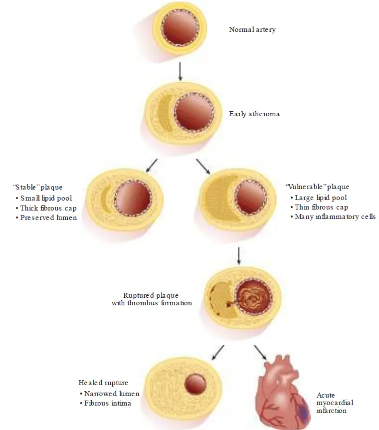
Despite the common use of these terms, this distinction vastly oversimplifies the heterogeneity of plaques and may overestimate the ability to foresee a plaque’s prognosis. For example, most plaques with the “vulnerable” morphology do not actually cause clinical events; hence, attempts to specifically identify such plaques may not direct therapy in an effective manner. Moreover, a substantial minority of fatal thrombi in coronary arteries arise from plaques with intact fibrous caps that may arise from superficial erosion of the lesion by mechanisms that are not well understood.
Rupture of atherosclerotic plaque does not inevitably cause major clinical events such as myocardial infarction or stroke. As described earlier, small non-occlusive thrombi may incorporate into the plaque, stimulating further smooth muscle growth and extracellular matrix deposition. The balance between the thrombogenic and fibrinolytic potential of the plaque, and the fluid phase of the blood determines whether disruption of the fibrous cap leads to a transient, nonobstructive mural thrombus or to a completely occlusive clot.
The probability of a major thrombotic event reflects the balance between the competing processes of clot formation and dissolution by fibrinolysis. Inflammatory stimuli found in the plaque microenvironment elicit tissue factor (which initiates the extrinsic coagulation pathway) and further support thrombosis by favoring the expression of anti-fibrinolytics (e.g. plasminogen activator inhibitor-1) over the expression of anticoagulants (e.g. thrombomodulin and heparin-like molecules) and pro-fibrinolytics (e.g. tissue plasminogen activator).
A person’s propensity toward coagulation may be enhanced by genetics, comorbid conditions (e.g. diabetes), and/or lifestyle factors (e.g., smoking, visceral obesity). Consequently, the concept of a “vulnerable plaque” has expanded to that of the “vulnerable patient,” to acknowledge other contributors to a person’s vascular risk.
Complications of Atherosclerosis
Atherosclerotic plaques do not distribute homogeneously throughout the vasculature. They usually develop first in the dorsal aspect of the abdominal aorta and proximal coronary arteries, followed by the popliteal arteries, descending thoracic aorta, internal carotid arteries, and renal arteries. Therefore, the regions perfused by these vessels most commonly suffer the consequences of atherosclerosis.
Atherosclerosis risk factors
In the early 20th century, most viewed atherosclerosis as an inevitable process of aging. But in 1948, the landmark Framingham Heart Study began to examine the relationship between specific attributes and cardiovascular disease, establishing the concept of atherosclerotic risk factors. Among later studies, the Multiple Risk Factor Intervention Trial (MRFIT) screened more than 325,000 men, offering an opportunity to correlate risk factors with subsequent cardiovascular disease.
Of the major risk factors, those that are not correctable include advanced age, male gender, and heredity (i.e. a history of coronary heart disease among first-degree relatives at a young age). Modifiable risk factors include dyslipidemia, tobacco smoking, hypertension, diabetes mellitus, and lack of physical activity and obesity.
Note
Young age implies before 55 years for a male relative or before 65 years for a female relative
In addition to these standard predictors, certain biologic markers associated with the development of cardiovascular events have been undergoing rigorous evaluation as “novel” risk markers. These include elevated circulating levels of the special lipoprotein particle Lp(a) and certain markers of inflammation, including the acute-phase reactant C-reactive protein (CRP).
Genetics
While directly causative genes remain elusive, recent genome-wide association studies (GWAS) have identified a number of loci associated with atherosclerosis. The strongest connection with myocardial infarction localizes to chromosome 9p21.3. This region contains genes that code for two cyclin-dependent kinase inhibitors that can regulate the cell cycle and may participate in TGF-β inhibitory pathways.
Other associations with coronary artery disease include SORT-1 that encodes a molecule implicated in lipoprotein trafficking.
Genetic studies have also shown that loss of function mutations in the gene that encodes the enzyme PCSK9 (proprotein convertase subtilisin/ kexin type 9) augment LDL receptor levels on cell surfaces, boosting LDL clearance, and yielding lower LDL concentrations in blood. Individuals with this mutation are thus exposed to lower levels of LDL than those with the typical genotype, and appear protected from atherosclerotic events. This observation has spurred the ongoing development of biological agents that limit PCSK9 action.
Dyslipidemia
A large and consistent body of evidence established abnormal circulating lipid levels as a major risk factor for atherosclerosis. Observational studies have shown that societies with high consumption of saturated fat and prevalent hypercholesterolemia have greater mortality from coronary disease than countries with traditionally low saturated fat intake and low serum cholesterol levels (e.g., rural Japan and certain Mediterranean nations).
Similarly, data from the Framingham Heart Study and other cohorts have shown that the risk of ischemic heart disease increases with higher total serum cholesterol levels. The coronary risk is approximately twice as high for a person with a total cholesterol level of 240 mg/ dL compared with a person whose cholesterol level is 200 mg/ dL.
When present in excess, LDL can accumulate in the subendothelial space and undergo the chemical modifications that further damage the intima, as described earlier, initiating the development of atherosclerotic lesions. Thus, LDL is commonly known as “bad cholesterol.” Conversely, elevated HDL particles (often called “good cholesterol”) associate with protection against atherosclerosis, often attributed to its ability to transport cholesterol away from the peripheral tissues back to the liver for disposal (termed “reverse cholesterol transport”) and its antioxidative and anti-inflammatory properties.
Elevated serum LDL may persist for many reasons, including a diet or abnormalities in the LDL receptor. Patients with genetic defects in the LDL receptor, which leads to a condition known as familial hypercholesterolemia, cannot remove LDL from the circulation efficiently. Heterozygotes of the LDL receptor defect display high plasma LDL levels and develop premature atherosclerosis. Homozygotes who completely lack functional LDL receptors may experience vascular events as early as the first decade of life.
Increasing evidence also implicates triglyceride-rich lipoproteins, such as VLDL and IDL, in the development of atherosclerosis. However, it remains undetermined whether these particles participate directly in the disease or simply keep company with low levels of HDL cholesterol.
Tobacco Smoking
Numerous studies have shown that tobacco smoking predisposes to atherosclerosis and ischemic heart disease. Even low level smoking leads to adverse outcomes, but the heaviest smokers have the greatest risk of cardiovascular events.
Tobacco smoking could promote atherosclerotic disease in several ways, including enhanced oxidative modification of LDL, decreased circulating HDL levels, endothelial dysfunction owing to tissue hypoxia and increased oxidant stress, increased platelet adhesiveness, inappropriate stimulation of the sympathetic nervous system by nicotine, and displacement of oxygen by carbon monoxide from hemoglobin. Extrapolation from animal experiments suggests that smoking not only accelerates atherogenesis but also increases the propensity for thrombosis.
Fortunately, smoking cessation can reverse some of the adverse outcomes. People who stop smoking greatly reduce their likelihood of coronary heart disease, compared with those who continue to smoke. In one study, after 3 years of cessation, the risk of coronary artery disease for former smokers became similar to subjects who never smoked.
Hypertension
The association between elevated blood pressure and cardiovascular disease does not appear to have a specific threshold. Rather, risk increases continuously with progressively higher pressure values. Systolic pressure predicts adverse outcomes more reliably than does diastolic pressure, particularly in older persons.
Hypertension may accelerate atherosclerosis in several ways. Animal studies have shown that elevated blood pressure injures vascular endothelium and may increase the permeability of the vessel wall to lipoproteins. Hypertension can enhance smooth muscle cell production of proteoglycans that bind and retain LDL particles, promoting their accumulation in the intima and facilitating their oxidative modification.
Angiotensin II, a mediator of hypertension, acts not only as a vasoconstrictor but also as a stimulator of oxidative stress (through activation of NADPH oxidase). Thus, hypertension may also promote atherogenesis by contributing to a prooxidant and inflammatory state.
Diabetes Mellitus and “Metabolic Syndrome”
With a three- to five- fold increased risk of acute coronary events, 80% of diabetic patients succumb to atherosclerosis-related conditions, including coronary heart disease, stroke, and peripheral artery disease.
The predisposition of diabetic patients to atherosclerosis may relate in part to accompanying dyslipidemia, to glycation of lipoproteins, or to the associated prothrombotic tendency. Diabetics frequently have impaired endothelial function, gauged by the reduced bioavailability of NO, and increased leukocyte adhesion.
Tight glycemic control in diabetic patients reduces the risk of microvascular complications, such as retinopathy and nephropathy. Yet demonstration of a reduction of macrovascular outcomes, such as myocardial infarction and stroke, by glycemic control remains much more elusive. In contrast to the uncertain benefits of intense glycemic control for macrovascular events, treatment of hypertension and dyslipidemia in diabetic patients convincingly reduces the risk of cardiac and cerebrovascular complications.
The metabolic syndrome (also known as “insulin resistance syndrome”) refers to a cluster of risk factors, including hypertension, hypertriglyceridemia, reduced HDL, hyperglycemia, and visceral obesity. This constellation associates with a high risk or atherosclerosis in both diabetic and nondiabetic patients. The presence of insulin resistance in this syndrome appears to promote atherogenesis long before affected persons develop overt diabetes.
Lack of Physical Activity
Exercise may mitigate atherogenesis in several ways. In addition to its beneficial effects on the lipid profile and blood pressure, exercise enhances insulin sensitivity and endothelial production of NO.
Estrogen Status
Before menopause, women have a lower incidence of coronary events than men. After menopause, however, men and women have similar rates. This observation suggests that estrogen may have atheroprotective properties. Physiologic estrogen levels in premenopausal women raise HDL and lower LDL. Experimentally, estrogen also exhibits potentially beneficial antioxidant and antiplatelet actions and improves endothelium-dependent vasodilation.
Early observational studies suggested that hormone therapy reduced the risk of coronary artery disease in postmenopausal women, prompting many physicians to prescribe such medications for cardiovascular prevention purposes. However, the Heart and Estrogen/ Progestin Replacement Study demonstrated an association between such hormone use and an early increased risk of vascular events in women with preexisting coronary disease. Subsequent randomized primary prevention studies from the Women’s Health Initiative were terminated prematurely because estrogen-plus-progestin treatment increased cardiovascular risk by 24% overall, with a striking 81% higher risk during the first year of therapy. Because currently available clinical trial data do not show that hormone therapy lowers cardiovascular events and that it may actually be harmful, such therapy should not be started for the sole goal of reducing cardiovascular risk.
See also
References
Additional Reading
- Cholesterol Treatment Trialists’ (CTT) Collaborators. Efficacy and safety of LDL-lowering therapy among men and women: meta-analysis of individual data from 174 000 participants in 27 randomised trials. Lancet. 2015;385:1397–1405.
- Cook NR, Paynter NP, Eaton CB, et al. Comparison of the Framingham and Reynolds risk scores for global cardiovascular risk prediction in the multiethnic women’s health initiative. Circulation. 2012;125:1748–1756.
- Gimbrone MA, Jr., García-Cardeña G. Vascular endothelium, hemodynamics, and the pathobiology of atherosclerosis. Cardiovasc Pathol. 2013;22:9–15.
- Bornfeldt KE, Tabas I. Insulin resistance, hyperglycemia, and atherosclerosis. Cell Metab. 2011;14:575–585.
- Libby P. Inflammation in atherosclerosis. Arterioscler Thromb Vasc Biol. 2012;32:2045–2051.
- Libby P. Mechanisms of acute coronary syndromes. N Engl J Med. 2013;369:883–884.
- Libby P, Ridker PM, Hansson GK. Progress and challenges in translating the biology of atherosclerosis. Nature. 2011;473:317–325.
- Moore KJ, Tabas I. Macrophages in the pathogenesis of atherosclerosis. Cell. 2011;145:341–355.
- Schunkert H, et al. Large-scale association analysis identifies 13 new susceptibility loci for coronary artery disease. Nat Genet. 2011;43:333–338.
- Stein EA, Mellis S, Yancopoulos GD, et al. Effect of a monoclonal antibody to PCSK9 on LDL cholesterol. N Engl J Med. 2012;366:1108–1118.
- Steinberg D. In celebration of the 100th anniversary of the lipid hypothesis of atherosclerosis. J Lipid Res. 2013;54:2946–2949.
- Stone NJ, Robinson J, Lichtenstein AH, et al. 2013 ACC/AHA guideline on the treatment of blood cholesterol to reduce atherosclerotic cardiovascular risk in adults. Circulation. 2013;129:S1–S45. DOI: 10.1161/01.cir.0000437738.63853.7a.
- Tsimikas S, Hall JL. Lipoprotein(a) as a potential causal genetic risk factor of cardiovascular disease: A rationale for increased efforts to understand its pathophysiology and develop targeted therapies. J Am Coll Cardiol. 2012;60:716–721.