Part 5: Ischemic Heart Disease
Tue Jul 21 2026
By B. Hassan
Introduction
The clinical presentation of ischemic heart disease can be highly variable and forms a spectrum of syndromes. For example, ischemia may be accompanied by uncomfortable sensation in the chest during exertion that subsides on rest. In other cases, it may occur without any clinical manifestations at all, a condition termed silent ischemia.
Angina pectoris remains the most common manifestation of ischemic heart disease and literally means “strangling in the chest.” Although other conditions may lead to similar discomfort, angina refers specifically to the uncomfortable sensation in the chest and neighboring structures that arises from an imbalance between myocardial oxygen supply and demand.
In the normal heart, the oxygen requirements of the myocardium match the coronary arterial supply. Even during vigorous exercise, when the metabolic needs of the heart increase, so does the delivery of oxygen to myocardial cells such that the balance is maintained. The mechanism behind this regulation is described over the following segments.
Myocardial Oxygen Supply
The supply of oxygen to the myocardium depends on the oxygen content of the blood and the rate of coronary blood flow. The oxygen content is determined by the hemoglobin concentration and the degree of systemic oxygenation. In the absence of anemia or lung disease, oxygen content remains fairly constant. In contrast, coronary blood flow is more dynamic, and regulation of that flow is responsible for matching the oxygen supply with metabolic requirements.
As in all blood vessels, coronary artery flow () is directly proportional to the vessel’s perfusion pressure () and is inversely proportional to coronary vascular resistance, that is:
However, unlike other arterial systems in which the greatest blood flow occurs during systole, the main coronary perfusion takes place during diastole. This is because systolic flow is impaired by the compression of the small coronary branches as they course through the contracting myocardium (mainly in the subendocardial region). On the other hand, coronary flow is unimpaired in diastole as the relaxed myocardium does not compress the coronary vasculature.
Thus, in coronary arteries, perfusion pressure can be approximated by the aortic diastolic pressure. Conditions that decrease aortic diastolic pressure (such as hypotension or aortic valve regurgitation) decrease coronary artery perfusion pressure and may lessen myocardial oxygen supply.
Coronary vascular resistance is the other determinant of coronary blood flow. This resistance is dynamically controlled by (1) forces that externally compress the coronary arteries and (2) factors that alter intrinsic coronary tone.
External compression is exerted on the coronary vessels during the cardiac cycle by contraction of the surrounding myocardium.
The degree of external coronary compression is directly related to intra-myocardial pressure and is therefore greatest during systole, as described earlier. Moreover, when the myocardium contracts, the subendocardium, adjacent to the high intraventricular pressure, is subjected to forces greater than the outer muscle layers. This is one reason the subendocardium is the region most vulnerable to ischemic damage.
Intrinsic Control of Coronary Arterial Tone
Unlike most tissues, the heart cannot increase oxygen extraction on demand because in its basal state, it removes nearly as much oxygen as possible from its blood supply. Thus, any additional oxygen requirement must be met by an increase in blood flow, and autoregulation of coronary vascular resistance is the most important mediator of this process.
Metabolites
The accumulation of local metabolites significantly affects coronary vascular tone and acts to modulate myocardial oxygen supply to meet changing metabolic demands. During hypoxia, aerobic metabolism and oxidative phosphorylation in the mitochondria are inhibited and generation of ATP is impaired. Consequently, adenosine diphosphate (ADP) and adenosine monophosphate (AMP) accumulate and are subsequently degraded to adenosine. Adenosine is a potent vasodilator and is thought to be the prime metabolic mediator of vascular tone. By binding to receptors on vascular smooth muscle, adenosine decreases calcium entry into cells, which leads to relaxation, vasodilatation, and increased coronary blood flow.
Other metabolites that act locally as vasodilators include lactate, acetate, hydrogen ions, and carbon dioxide. These are also increased in hypoxic states, leading to further vasodilation, aiding in the autoregulation process.
Endothelial Factors
Arterial endothelial cells produce numerous vasoactive substances that contribute to the regulation of vascular tone as described in the previous part talking about atherosclerosis. Normal endothelial cells synthesize potent vasodilator substances that contribute to the modulation of vascular tone. Among the first of these to be identified were prostacyclin (an arachidonic acid metabolite) and the nitric oxide (NO) radical.
In experimental preparations, it was shown that acetylcholine (ACh) has two opposite actions on blood vessels. Its direct effect on vascular smooth muscle cells is to cause vasoconstriction, but when an intact endothelial lining overlies the smooth muscle cells, vasodilatation occurs instead. Subsequent experiments showed that ACh causes the endothelial cells to release NO, which quickly diffuses to the adjacent smooth muscle cells causing vasodilatation.
Note
NO induces vasodilation by activating the enzyme guanylyl cyclase, which produces cyclic guanosine monophosphate (cGMP), which results in smooth muscle relaxation through a reduction in cytosolic . On the other hand, prostacyclin induces vasodilation by a cyclic AMP–dependent mechanism.
Intracoronary administration of ACh in a normal person causes vasodilatation, presumably through the release of NO. However, in conditions associated with endothelial dysfunction (e.g. atherosclerosis), intracoronary ACh administration results in paradoxical vasoconstriction instead. This likely reflects reduced production of NO by the dysfunctional endothelial cells, resulting in unopposed direct vasoconstriction by ACh. Of particular interest is that the loss of vasodilatory response to ACh is evident in patients with certain cardiac risk factors (e.g. hypercholesterolemia, hypertension, cigarette smoking) even before the appearance of atheromatous plaque. Thus, the impaired release of NO may be an early and sensitive predictor for the later development of atherosclerotic lesions.
In contrast to the endothelial-dependent vasodilators, some agents cause smooth muscle relaxation independent of the presence of endothelial cells. For example, the drugs sodium nitroprusside and nitroglycerin result in vasodilatation by providing an exogenous source of NO, forming cGMP without endothelial cell participation.
Other factors like hypoxia, shear stress, and platelet products (e.g., serotonin) also induce release of vasodilator substances like NO and prostacyclin. Many of these also have dual actions just like ACh.
Endothelium-derived hyperpolarizing factor (EDHF) also appears to have important vasodilatory properties. Like NO, it is a diffusible substance released by the endothelium that causes hyperpolarization (i.e. relaxation) of vascular smooth muscle cells. EDHF is released by some of the same factors that stimulate NO, including ACh and normal pulsatile blood flow. In the coronary circulation, EDHF appears to be more important in modulating relaxation in small arterioles than in the large arteries.
Under normal circumstances, the healthy endothelium promotes vascular smooth muscle relaxation (vasodilatation) through elaboration of substances such as NO and prostacyclin, which predominate over the endothelial vasoconstrictors. However, dysfunctional endothelium (e.g. in atherosclerotic vessels) secretes reduced amounts of vasodilators, causing the balance to shift toward vasoconstriction instead.
Neural Factors
The neural control of vascular resistance has both sympathetic and parasympathetic components. Under normal circumstances, the contribution of the parasympathetic nervous system appears minor, but sympathetic receptors play an important role. Coronary vessels contain both α-adrenergic and β2-adrenergic receptors. Stimulation of α-adrenergic receptors results in vasoconstriction, whereas β2-receptors promote vasodilatation.
It is the interplay among the metabolic, endothelial, and neural regulating factors that determines the net impact on coronary vascular tone. For example, catecholamine stimulation of the heart may initially cause coronary vasoconstriction via the α-adrenergic receptor neural effect. However, catecholamine stimulation also increases myocardial oxygen consumption through increased heart rate and contractility (β1-adrenergic effect), and the resulting increased production of metabolites induces net coronary dilatation instead.
Myocardial Oxygen Demand
The 3 major determinants of myocardial oxygen demand are (1) ventricular wall stress, (2) heart rate, and (3) contractility (which is also termed the inotropic state). Additionally, very small amounts of oxygen are consumed in providing energy for basal cardiac metabolism and electrical depolarization.
Ventricular wall stress () is the tangential force acting on the myocardial fibers, tending to pull them apart, and energy is expended in opposing that force. Wall stress is related to intraventricular pressure (), the radius of the ventricle (), and ventricular wall thickness () and is approximated by Laplace’s relationship:
This means that wall stress is directly proportional to systolic ventricular pressure. Circumstances that increase pressure in the left ventricle, such as aortic stenosis or hypertension, increase wall stress and myocardial oxygen consumption. Conditions that decrease ventricular pressure, such as antihypertensive therapy, reduce myocardial oxygen consumption.
Because wall stress is also directly proportional to the radius of the left ventricle, conditions that increase LV filling (e.g. mitral or aortic regurgitation) raise wall stress and oxygen consumption. Conversely, any physiologic or pharmacologic maneuver that decreases LV filling and size (e.g., nitrate therapy) reduces wall stress and myocardial oxygen consumption.
Finally, wall stress is inversely proportional to ventricular wall thickness because the force is spread over a greater muscle mass. A hypertrophied heart has lower wall stress and oxygen consumption per gram of tissue than a thinned-walled heart. Thus, when hypertrophy develops in conditions of chronic pressure overload, such as aortic stenosis, it serves a compensatory role in reducing oxygen consumption.
Note
Although hypertrophy lowers wall stress per unit mass, total myocardial oxygen demand in hypertrophied states is actually increased overall due to massive total mass and high systolic pressure.
The second major determinant of myocardial oxygen demand is heart rate. If the heart rate accelerates, the number of contractions and the amount of ATP consumed per minute increases and oxygen requirements increase. Conversely, slowing the heart rate (e.g., with a β-blocker drug) decreases ATP utilization and oxygen consumption. Of note about heart rate is that tachycardia mainly reduces the time of diastole in each cardiac cycle (i.e. the time most important for coronary perfusion), which can be detrimental for myocardial ischemia.
The third major determinant of oxygen demand is myocardial contractility. Circulating catecholamines, or the administration of positive inotropic drugs, directly increase the force of contraction, which increases oxygen utilization. Conversely, negative inotropic effectors, such as β-adrenergic–blocking drugs, decrease myocardial oxygen consumption.
In the normal state, autoregulatory mechanisms adjust coronary tone to match oxygen supply with oxygen requirements. In the absence of obstructive coronary disease, these mechanisms maintain a fairly constant coronary flow rate, as long as the aortic perfusion pressure is approximately 60 mmHg or greater. In the setting of advanced coronary atherosclerosis, however, the fall in perfusion pressure distal to the arterial stenosis, along with dysfunction of the endothelium in the involved segment causes a mismatch between the available blood supply and myocardial metabolic demands.
Pathophysiology of ischemia
Myocardial ischemia in coronary artery disease results from the combination of fixed vessel narrowing and abnormal vascular tone, contributed to by atherosclerosis-induced endothelial cell dysfunction.
Fixed Vessel Narrowing
Poiseuille’s law states that for flow through a vessel:
in which
- is flow
- is the pressure difference between the points being measured
- is the vessel radius
- is the fluid viscosity
- is the vessel length
And since flow is also equal to the pressure difference divided by the resistance (R) to flow:
By combining these two formulas and rearranging, resistance to blood flow in a vessel can be expressed as:
Thus, vascular resistance is governed, in part, by the length of the vessel, and more importantly, by the degree of vessel narrowing.
Note
The length of the vessel is considered “constant” as the length of blood vessels don’t change much throughout life. Vessel radius is the most important factor for determining blood flow through a vessel, as it is a dynamic quantity that can change (i.e. vasodilation or vasoconstriction). Moreover, a doubling of the vessel length only increases vascular resistance by a factor of 2, while halving of the radius of that vessel increases the resistance by a factor of 16. Thus when talking about vascular resistance, we usually only consider the vessel radius.
The coronary arteries consist of large, proximal epicardial segments and smaller, distal arterioles. The proximal vessels are subject to overt atherosclerosis that results in stenotic plaques. The distal arterioles are usually free of stenotic plaques and can adjust their vasomotor tone in response to metabolic needs. These arterioles serve as “dams”, increasing their diameter with exertion to meet increasing oxygen demand.
The hemodynamic significance of a coronary artery narrowing depends on both the degree of stenosis of the epicardial portion of the vessel and the amount of compensatory vasodilatation the distal resistance vessels are able to provide:
-
If a stenosis narrows the lumen diameter by less than 60%, the maximal potential blood flow through the artery is not significantly altered and, in response to exertion, arterioles can dilate to provide adequate blood flow.
-
If the stenosis narrows the diameter by more than 70%, resting blood flow is normal, but maximal blood flow is reduced even with full dilatation of arterioles. In such situations, when oxygen demand increases (e.g. due to physical exertion), coronary flow is inadequate, and oxygen demand exceeds supply, and myocardial ischemia results.
-
If the stenosis compromises the vessel lumen by more than 90%, then even with maximal dilatation of arterioles, blood flow may be inadequate to meet basal requirements and ischemia can develop at rest.
Note
Although collateral circulation at sites distal to atherosclerotic stenoses may buffer the fall in myocardial oxygen supply, it is often not sufficient to prevent ischemia during exertion in critically narrowed vessels (i.e. angina).
Endothelial Dysfunction
In addition to fixed vessel narrowing, the other major contributor to reduced myocardial oxygen supply in chronic coronary artery disease is endothelial dysfunction. Abnormal endothelial function can contribute to the ischemia in two ways:
- Inappropriate vasoconstriction of coronary arteries
- Loss of normal antithrombotic properties
Normally, physical activity or mental stress results in measurable coronary artery vasodilatation. This effect is thought to be regulated by the sympathetic nervous system and the release of vasoactive substances as discussed earlier. However, in patients with dysfunctional endothelium (e.g. atherosclerosis), an impaired release of endothelial vasodilators leaves the direct catecholamine effect unopposed, such that vasoconstriction occurs instead. Moreover, the vasodilatory effect of local metabolites is attenuated if the endothelium is dysfunctional, further uncoupling the regulation of vascular tone from metabolic demands.
In addition to their vasodilatory action, factors released from endothelial cells (e.g. NO and prostacyclin) also exert anti-thrombotic properties by interfering with platelet aggregation. However, in endothelial dysfunction, release of these substances is impaired; thus, the anti-thrombotic effect is attenuated. Thus, in syndromes characterized by thrombosis (i.e. acute coronary syndromes), the impaired release of NO and prostacyclin allows platelets to aggregate and to secrete their procoagulants and vasoconstrictors.
Consequences of ischemia
The consequences of ischemia reflect the inadequate myocardial oxygenation and local accumulation of metabolites. For example, during ischemia, myocytes convert from aerobic to anaerobic metabolic pathways. The reduced ATP impairs the contractile proteins and results in a transient reduction of both ventricular systolic contraction and diastolic relaxation, as each are energy-dependent processes.
The consequent elevation of LV diastolic pressure is transmitted (via the left atrium and pulmonary veins) to the pulmonary capillaries and can precipitate pulmonary congestion and the symptom of dyspnea.
In addition, metabolites like lactate, serotonin, and adenosine accumulate locally. It is suspected that one or more of these metabolites activate peripheral pain receptors in the C7 through T4 distribution and may be the mechanism by which the discomfort of angina is produced.
The accumulation of local metabolites and transient abnormalities of myocyte ion transport may also precipitate arrhythmias.
The ultimate fate of ischemic myocardium depends on the severity and duration of the imbalance between oxygen supply and demand. It was previously thought that ischemic cardiac injury results in either irreversible myocardial necrosis (i.e. MI) or rapid and full recovery of myocyte function. It is now known that in addition to those outcomes, ischemia can sometimes result in a period of prolonged contractile dysfunction without myocyte necrosis, and recovery of normal function may ultimately follow.
For example, stunned myocardium refers to tissue that, after suffering an acute episode of severe ischemia (but not necrosis), demonstrates prolonged systolic dysfunction even after the return of normal blood flow. In this setting, the abnormalities following ischemia are reversible without coronary intervention and contractile function recovers over days to weeks once blood flow is back. The mechanism responsible for this delayed recovery of function involves myocyte calcium overload and the accumulation of oxygen-derived free radicals. In general, the magnitude of stunning is proportional to the degree of the preceding ischemia, and this state is likely the consequence of an ischemic episode that just falls short of causing irreversible necrosis.
In contrast, hibernating myocardium refers to tissue that undergoes chronic contractile dysfunction due to a persistently reduced blood supply. In this situation, irreversible damage has not occurred and ventricular function can improve if appropriate blood flow is restored, though it usually requires percutaneous or surgical revascularization. The mechanism responsible for this state is the adaptive myocardial response to the chronic ischemia, which downregulates its metabolic needs and contractility to match the reduced supply.
Special imaging studies (e.g. PET scan or dobutamine echocardiography) can differentiate hibernating from infarcted myocardium. That distinction can help guide the decision of whether to undergo coronary revascularization, because hibernating myocardium would be expected to regain contractile function with restoration of blood flow, whereas infarcted myocardium would not.
Stable Angina
Stable angina manifests as a pattern of transient chest discomfort during exertion or emotional stress. It is generally caused by fixed, obstructive atheromatous plaque in one or more coronary arteries. The severity of symptoms is usually related to the degree of stenosis (i.e. the percentage of coronary artery lumen obstruction) as discussed earlier.
Potentially contributing to the inadequate oxygen supply in stable angina is inappropriate coronary vasoconstriction caused by atherosclerosis-associated endothelial dysfunction.
As a result, the extent of coronary artery narrowing in patients with atherosclerosis is not necessarily constant. Rather, it can vary from moment to moment because of changes in the coronary vascular tone. For some patients with stable angina, alterations in tone play a minimal role in the decreased myocardial oxygen supply, and the level of exertion required to precipitate angina is fairly constant. These patients have fixed-threshold angina. In other cases, the degree of obstruction caused by vasoconstriction or vasospasm plays a more prominent role, and such patients may have variable-threshold angina.
For example, on a given day, a patient with variable-threshold angina can exert themselves without chest discomfort, but on another day, the same degree of exertion produces symptoms. The difference reflects alterations in vascular tone over the sites of fixed stenosis.
Unstable Angina
A patient with chronic stable angina may experience a sudden increase in the frequency or duration of ischemic episodes, occurring with lesser degrees of exertion and even at rest. This acceleration of symptoms is known as unstable angina, which can be a precursor to an acute myocardial infarction.
Unstable angina and acute myocardial infarction are also known as acute coronary syndromes and result from various pathophysiologic mechanisms, most commonly rupture of an unstable atherosclerotic plaque with subsequent platelet aggregation and thrombosis.
Variant Angina
A small minority of patients present with episodes of local coronary artery spasm in the absence of atherosclerotic lesions. This syndrome is known as variant angina or Prinzmetal angina.
In this case, intense vasospasm alone reduces coronary oxygen supply and results in angina. The mechanism by which such profound spasm develops is not completely understood but may involve increased sympathetic activity in combination with endothelial dysfunction. It is thought that many patients with variant angina may actually have early atherosclerosis manifested only by a dysfunctional endothelium.
Variant angina often occurs at rest because the ischemia results from transient reduction of the coronary oxygen supply rather than an increase in myocardial oxygen demand.
Note
Unlike variable-threshold stable angina, variant angina is not related to effort.
Silent Ischemia
Episodes of cardiac ischemia sometimes occur in the absence of perceptible discomfort or pain, such instances are referred to as silent ischemia. These asymptomatic episodes can occur in patients who experience typical symptomatic angina. Conversely, in some patients, silent ischemia may be the only manifestation of coronary artery disease.
It may be difficult to diagnose silent ischemia clinically, but its presence can be detected by laboratory techniques such as continuous ambulatory electrocardiography or it can be elicited by exercise stress testing.
One study estimated that silent ischemic episodes occur in 40% of patients with stable angina and in 2.5% to 10% of asymptomatic middle-aged men.
The reason why some episodes of ischemia are silent whereas others are symptomatic has not been elucidated. The degree of ischemia cannot fully explain the disparity, because even myocardial infarction may present without symptoms in some patients. Silent ischemia has been reported to be more common among diabetic patients (possibly due to peripheral neuropathy), the elderly, and in women.
Syndrome X
The term syndrome X refers to patients with typical symptoms of angina pectoris who have no evidence of significant atherosclerotic coronary stenoses on coronary angiograms. Some of these patients may show definite laboratory signs of ischemia during exercise testing.
The pathogenesis of ischemia in this situation may be related to inadequate vasodilator reserve of the coronary arteriole. It is thought that the arterioles (which are too small to be visualized by coronary angiography) may not dilate appropriately during periods of increased myocardial oxygen demand.
Patients with syndrome X have a better prognosis than those with overt atherosclerotic disease.
Clinical features of angina
The most important part of the clinical evaluation of ischemic heart disease is the history described by the patient. Because chest pain is a common complaint, it is important to focus on the characteristics that help distinguish myocardial ischemia from other causes of chest discomfort.
Angina is most often described as a “pressure,” “discomfort,” “tightness,” or “heaviness” in the chest. It is rare that the sensation is actually described as a “pain,” and often a patient will correct the physician who refers to the symptoms as “pain”. Sometimes, a patient describes the sensation as “an elephant sitting on my chest.” Anginal discomfort is neither sharp nor stabbing, and it does not vary with movement of the chest wall. It is a steady discomfort that lasts a few minutes, rarely more than 5 to 10 minutes. It always lasts more than a few seconds. This helps differentiate it from sharper and briefer musculoskeletal pains.
While describing angina, the patient may place a clenched fist over their sternum, referred to as Levine sign, as if defining the constricting discomfort by that tight grip.
Angina is usually diffuse rather than localized to a single point. It is most often located in the retrosternal area or in the left precordium but may occur anywhere in the chest, back, arms, neck, lower face, or upper abdomen. It often radiates to the shoulders and inner aspect of the arms, especially on the left side.
Ischemia also results in transient dysfunction of LV systolic contraction and diastolic relaxation. The resultant elevation of LV diastolic pressure is transmitted to the pulmonary vasculature and often causes dyspnea during the episode. When such symptoms occur due to myocardial ischemia but are unaccompanied by typical chest discomfort, they are referred to as “anginal equivalents.”
Angina, when not caused by pure vasospasm, is precipitated by conditions that increase myocardial oxygen demand, mainly physical exertion or emotional stress. Additional factors that increase myocardial oxygen demand and can precipitate anginal discomfort include a large meal or cold weather. The latter induces peripheral vasoconstriction, which in turn augments myocardial wall stress as the left ventricle contracts against the increased resistance.
Angina is generally relieved within minutes after cessation of the activity that precipitated it and even more quickly (within 3 to 5 minutes) by sublingual nitroglycerin. This response can help differentiate myocardial ischemia from many of the other conditions that produce chest discomfort. Patients with variant angina often develop symptoms at rest, independent of activities that increase myocardial oxygen demand.
Although the level of exertion necessary to precipitate angina may remain constant, the frequency of episodes varies considerably because patients quickly learn which activities cause their symptoms and avoid them. It is thus important to inquire about reductions in activities of daily living when taking the history.
If it is possible to examine a patient during an anginal attack, several transient physical signs may be detected. Tachycardia, diaphoresis, and hypertension are common because of the augmented sympathetic response. Myocardial ischemia may lead to papillary muscle dysfunction and therefore mitral regurgitation. Ischemia decreases ventricular compliance, producing a stiffened ventricle and therefore an S4 gallop.
Differential Diagnosis
Several conditions can give rise to symptoms that mimic the chest discomfort of angina pectoris. In contrast to angina pectoris, gastrointestinal causes of recurrent chest pain are often precipitated by certain foods and are unrelated to exertion. Musculoskeletal causes of chest discomfort tend to be more superficial or can be localized to a discrete spot (i.e., the patient can point to the pain with one finger). Similarly, the presence of pleuritic pain (sharp pain aggravated by respiratory movements) argues against angina as the cause; this symptom is more likely a result of pericarditis, or an acute pulmonary condition such as pulmonary embolism or acute pneumothorax.
The following are the common causes of recurrent chest pain and how to differentiate them from angina pectoris.
Pericarditis:
- Sharp, pleuritic pain that varies with position
- Friction rub may be present on auscultation
- Can last for hours to days
- ECG shows diffuse ST elevations and PR deviation
Gastroesophageal reflux:
- Retrosternal burning pain
- Precipitated by certain foods
- Worsened by supine position
- Unaffected by exertion
- Relieved by antacids
Peptic ulcer disease:
- Epigastric ache or burning
- Occurs after meals, unaffected by exertion
- Relieved by antacids
Esophageal spasm:
- Retrosternal pain accompanied by dysphagia
- Precipitated by meals
- Unaffected by exertion
- May be relieved by nitroglycerin
Costochondral syndrome:
- Sternal pain worsened by chest movement
- Costochondral junctions tender to palpation
- Relieved by anti-inflammatory drugs, not by nitroglycerin
Cervical radiculitis:
- Constant ache or shooting pains
- May be in a dermatomal distribution
- Worsened by neck motion
Diagnostic studies
Once angina is suspected, several diagnostic studies may be helpful in confirming myocardial ischemia. Because many of these tests are costly, it is important to choose the appropriate studies for each patient.
ECG
An ECG obtained during an anginal episode is one of the most helpful tools for diagnosis of myocardial ischemia. Although it is easy to arrange when symptoms occur in hospitalized patients, it may not be possible to “catch” an episode in an outpatient basis.
Acute ischemia usually results in transient horizontal or downsloping ST depression and T-wave flattening or inversions.
Note
Horizontal ST depression greater than 1mm or a downsloping ST depression have a sensitivity of approximately 65% to 70% and a specificity of 75% to 80% for the detection of anatomically significant coronary artery disease.
Occasionally, ST-segment elevations are seen, suggesting more severe transmural myocardial ischemia, and can also be observed during variant angina.
In contrast to the ECG of a patient with acute MI, the ST deviations seen in patients with stable angina quickly normalize with resolution of the symptoms. In fact, ECGs obtained during periods free of ischemia are completely normal in approximately half of patients with stable angina. In others, “nondiagnostic” ST and T-wave deviations may be present.
Evidence of a previous MI (e.g. pathologic Q waves) on the ECG also points to the presence of underlying coronary disease.
Stress Testing
Because ECGs obtained between anginal episodes may be normal, such tracings do not rule out ischemic heart disease. For this reason, provocative exercise or pharmacologic stress tests are valuable diagnostic tools.
During this test, the patient exercises on a treadmill or a stationary bicycle with progressively higher workloads and is observed; the heart rate and ECG are continuously monitored, and blood pressure is checked at regular intervals. The test is continued until angina develops, signs of myocardial ischemia appear on ECG, a target heart rate is achieved (85% of the maximal predicted heart rate), or the patient becomes too fatigued to continue.
Note
The maximal predicted heart rate is calculated by subtracting the patient’s age from 220 bpm.
The test is considered abnormal if the patient’s typical chest discomfort is reproduced or if ECG abnormalities consistent with ischemia develop. The test is considered markedly positive if one or more of the following signs occur:
-
Ischemic ECG changes develop in the first 3 minutes of exercise or persist 5 minutes after exercise has stopped
-
The magnitude of the ST depression is greater than 2 mm
-
The systolic blood pressure abnormally falls during exercise, resulting from ischemia-induced impairment of contractile function
-
High-grade ventricular arrhythmias develop
-
The patient cannot exercise for at least 2 minutes
The results of a stress test may be affected by medications like β-blockers or calcium channel blockers, which may impair the ability to achieve the target heart rate. In these situations, one must consider the purpose of the stress test. If it is to determine whether ischemic heart disease is present, then those medications should be withheld for 1-2 days before the test. On the other hand, if the patient has known ischemic heart disease and the aim is to assess the efficacy of the current medical regimen, testing should be performed while the patient takes their usual medications.
Nuclear Imaging Studies
Since a standard stress test relies on ischemia-related changes on ECG, the test is less useful in patients with baseline ECG abnormalities (e.g. left bundle branch block or LV hypertrophy). In these situations, radionuclide imaging can be combined with exercise testing to overcome these limitations and to increase the sensitivity and specificity of the study.
During the test, a radionuclide (either a technetium-99m–labeled compound or thallium-201) is injected IV at peak exercise, after which imaging is performed. The radionuclide accumulates in proportion to the degree of perfusion of the myocardial cells. Areas of poor perfusion (i.e. ischemic regions) during exercise do not accumulate radionuclide and appear as “cold spots” on the image. However, irreversibly infarcted areas also do not take up the radionuclide, and they too will appear as cold spots.
To differentiate between transient ischemia and infarcted tissue, imaging is also performed at rest (either before or several hours after exercise). If the cold spot fills in at rest, it indicates transient ischemia. If the cold spot remains unchanged at rest, it points to irreversible infarction.
Standard radionuclide exercise tests are 80% to 90% sensitive and approximately 80% specific for the detection of clinically significant coronary artery disease. PET scans (another form of nuclear stress imaging) provide superior spatial and temporal resolution, with sensitivity and specificity of 90% or greater. However, PET scans are not as widely available, and are generally more expensive.
Because nuclear imaging techniques are expensive, their use in screening for coronary artery disease should be reserved for:
-
Patients with baseline ECG abnormalities that obscure the interpretation of a standard exercise test
-
Improvement in test sensitivity when standard stress test results are discordant with the clinical symptoms
Pharmacologic Stress Tests
Patients unable to exercise can undergo pharmacological stress testing instead of the standard exercise stress test. This test uses various agents, including vasodilators or inotropes. The most common approach is to use a coronary vasodilator such as adenosine, regadenoson, or dipyridamole. Administration of these agents is typically coupled with nuclear imaging to reveal regions of myocardial ischemia.
Adenosine and regadenoson bind to the adenosine receptors on vascular smooth muscle cells, resulting in coronary vasodilatation. As ischemic regions are already maximally dilated (in compensation for the epicardial coronary stenoses), the vasodilatation induced by these agents increases flow to the myocardium perfused by healthy coronary arteries and thus “steals” blood away from the diseased segments.
Dipyridamole causes a similar effect indirectly, as it blocks normal cellular uptake of adenosine, thereby increasing adenosine’s concentration and subsequent stimulation of the receptor.
An alternative to vasodilating agents is inotropes like dobutamine, which increases myocardial oxygen demand by increasing heart rate and contractility, simulating some of the effects of exercise.
Vasodilator pharmacologic stress testing is generally preferred over dobutamine as it has a better safety profile and is technically easier and faster to perform. However, the vasodilator agents can cause bronchospasm in patients with reactive airways disease (by stimulating bronchiolar adenosine receptors) and should be avoided in such patients. In addition, a vasodilator test cannot be performed successfully on a patient exposed to methylxanthines (e.g. caffeine consumption or the bronchodilator theophylline) on the day of the study, as such agents competitively antagonize adenosine’s interaction with its receptor and blunt its effect.
Coronary Angiography
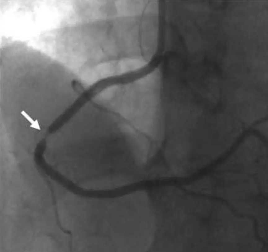
The most direct means of identifying coronary artery stenoses is by coronary angiography, in which atherosclerotic lesions are visualized radiographically following the injection of radiopaque contrast material. Although generally safe, the procedure is associated with a small risk of complications due to its invasive nature. Therefore, coronary angiography is typically reserved for patients whose anginal symptoms do not respond to pharmacologic therapy, or those with an unstable presentation, or when the results of noninvasive testing are so abnormal that severe coronary artery disease warranting revascularization is likely.
The following shows a coronary stenosis as visualized by contrast angiography:
When the degree of stenosis or its hemodynamic significance is unclear, additional techniques can be applied in the cardiac catheterization laboratory. For example, fractional flow reserve (FFR) measurement is a technique that can assess the functional severity of a stenosis identified at angiography. A special manometer-tipped guidewire inserted through the catheter measures the pressure in the coronary artery distal to the stenosis during induced vasodilatation. The FFR value is equal to the pressure distal to the stenosis () relative to the pressure proximal to the stenosis in the aorta ().
A higher FFR indicates a less severe stenosis. FFR less than 0.75 to 0.80 indicate severe stenoses that typically warrant mechanical intervention.
Although coronary angiography is considered the “gold standard” for the diagnosis of coronary artery disease, it should be noted that it provides only anatomic information. The clinical significance of lesions detected by angiography depends on both the degree of narrowing and also on the pathophysiologic consequences. Therefore, treatment decisions are made not only on the finding of stenoses but also by their effects, manifested by the patient’s symptoms, the viability of the myocardial segments served by stenotic vessels, and the degree of ventricular contractile dysfunction. Furthermore, standard arteriography does not reveal the composition of atherosclerotic plaque or its vulnerability to rupture.
Treatment
The goals of therapy in ischemic heart disease are to decrease the frequency of anginal attacks, to prevent acute coronary syndromes such as MI, and to prolong survival. A long-term crucial step is to address the risk factors that led to the development of atherosclerotic disease.
There is a good evidence to the benefit of smoking cessation, cholesterol improvement, and blood pressure control in lowering the risk of coronary disease events. Improvements in other risk factors for coronary artery disease, including serum glucose in diabetics, obesity and physical inactivity, may also reduce the risk of adverse outcomes although the benefits of these interventions are less well documented.
Treatment of an Acute Episode of Angina
When experiencing acute angina, the patient should cease physical activity. Sublingual nitroglycerin, an organic nitrate, is the drug of choice in this situation, which begins to take effect in 1 to 2 minutes. Nitrates relieve ischemia primarily through vascular smooth muscle relaxation, particularly venodilatation.
Venodilatation reduces venous return to the heart, decreasing LV volume and thus decreasing ventricular wall stress. This decreases myocardial oxygen consumption, thus helping restore oxygen balance in the ischemic heart.
A second action of nitrates is to dilate the coronary vasculature, with subsequent augmentation of coronary blood flow. This effect may be of little value in patients with angina in whom maximal coronary dilatation has already occurred due to accumulation of local metabolites. However, when coronary vasospasm plays a role in the development of ischemia, nitrate-induced coronary vasodilatation may be particularly beneficial.
Treatment to Reduce Frequency of Episodes
Pharmacologic agents can help decrease cardiac workload (i.e. reducing myocardial oxygen demand) and to increase myocardial perfusion. These are the first line in the prevention of anginal attacks. The three classes of medications most commonly used are β-adrenergic blockers, organic nitrates, and calcium channel blockers.
β-Blockers are directed against β-receptors, of which there are two classes: β1-adrenergic receptors are restricted to the myocardium, whereas β2-adrenergic receptors are located throughout the blood vessels and the bronchial tree. The stimulation of β1-receptors increases heart rate and contractility, increasing myocardial oxygen demand. Consequently, β-Blockers decrease ventricular contractility and heart rate, thereby relieving ischemia by reducing myocardial oxygen demand. In addition, slowing the heart rate may benefit myocardial oxygen supply by increasing the time spent in diastole, the phase when coronary perfusion primarily occurs.
In addition β-Blockers have been shown to decrease the incidence of MI, decrease recurrence of MI, and decrease mortality following an acute MI. Thus, β-blockers are first-line chronic therapy in the treatment of coronary artery disease.
β-Blockers are generally well tolerated but have several potential side effects. For example, they may precipitate bronchospasm in patients with underlying asthma by antagonizing β2-receptors in the bronchial tree. Although β1-selective blockers are theoretically less likely to exacerbate bronchospasm in such patients, drug selectivity for the β1-receptor is not complete, and in general, all β-blockers should be used cautiously, or avoided, in patients with significant obstructive lung disease. β-Blockers are also relatively contraindicated in patients with marked bradycardia or certain types of heart block to avoid additional impairment of electrical conduction
β-Blockers are also not used in patients with acutely decompensated LV dysfunction as they could intensify heart failure symptoms by further reducing contractility. However, they actually improve outcomes in patients with stable chronic heart failure. One might also expect that β-blockers would decrease myocardial blood perfusion by blocking the vasodilating β2-adrenergic receptors of the coronary arteries. However, this effect is usually attenuated by autoregulation and vasodilatation of the coronary vessels owing to the accumulation of metabolites.
β-Blockers sometimes cause fatigue and sexual dysfunction. They should be used with caution in diabetic patients because they can mask tachycardia and other symptoms that act as a warning signal for hypoglycemia.
Organic nitrates, as previously explained, relieve ischemia primarily through venodilatation and possibly through coronary vasodilatation. Sublingual nitroglycerin tablets or sprays are used in the treatment of acute attacks because of their rapid onset of action. In addition, they can be taken prophylactically before exertion to mitigate anginal attacks. Longer-acting anginal prevention can be achieved through a variety of nitrate preparations, including oral tablets of isosorbide dinitrate (or mononitrate) or a transdermal nitroglycerin patch. A limitation to chronic nitrate therapy is the development of drug tolerance, which occurs to some degree in most patients. Tolerance can be overcome by providing a nitrate-free interval for several hours each day, usually during sleep.
There is no evidence that nitrates improve survival or prevent infarctions, and they are used purely for symptomatic relief. Common side effects include headache and palpitations induced by vasodilatation and reflex tachycardia. The latter can be prevented by combining a β-blocker with the nitrate regimen.
Calcium channel blockers antagonize L-type calcium channels, but the actions of the individual drugs of this group vary. The dihydropyridines (e.g. nifedipine and amlodipine) are potent vasodilators. They relieve myocardial ischemia by causing venodilatation thus reducing oxygen demand (i.e. just like organic nitrates) and increasing myocardial oxygen supply via coronary dilatation. By the latter mechanism, they are also potent agents for the relief of coronary artery vasospasm.
Nondihydropyridine calcium channel blockers (verapamil and diltiazem) also act as vasodilators but are not as potent in this regard as the dihydropyridines. However, these have the additional cardiac depressant action, reducing ventricular contractility and slowing the heart rate, ultimately decreasing myocardial oxygen demand.
Questions have been raised about the safety of short-acting calcium channel–blocking drugs in the treatment of ischemic heart disease. In meta-analyses of randomized trials, these drugs have been associated with an increased incidence of MI and mortality. The adverse effect may relate to the rapid hemodynamic effects and blood pressure swings induced by the short-acting agents. Therefore, only long-acting calcium channel blockers are recommended for chronic angina, generally as second-line drugs if symptoms are not well controlled by β-blockers and nitrates.
Note
Care should be taken when combining a β-blocker with nondihydropyridine calcium channel blockers because the additive negative chronotropic effect can cause excessive bradycardia and the combined negative inotropic effect could precipitate heart failure.
Ranolazine, a 4th type of anti-ischemic therapy, has been shown to decrease the frequency of anginal attack and improve exercise tolerance in patients with chronic coronary artery disease. It differs from other anti-ischemic drugs in that it does not affect the heart rate or blood pressure. Although its mechanism of action is not fully understood, it is believed to inhibit the late phase of the action potential’s inward sodium current in ventricular myocytes. That late phase tends to be abnormally enhanced in ischemic myocardium, and the associated increased sodium influx results in higher-than-normal intracellular (mediated by the exchanger). Such calcium overload is thought to result in impaired diastolic relaxation and contractile insufficiency. Studies have supported ranolazine’s effectiveness in reducing angina, and its long-term safety, when used alone or in combination with other antianginal drugs.
Treatment to Prevent Acute Cardiac Events
Platelet aggregation and thrombosis are key elements in the pathophysiology of acute MI and unstable angina. Antiplatelet therapy reduces the risk of these acute coronary syndromes in patients with chronic angina and should be a standard part of their medication regimen. For example, aspirin has antithrombotic actions by inhibiting the synthesis of thromboxane A2, a mediator of platelet activation, as well as anti-inflammatory properties that may be important in stabilizing atheromatous plaque. Unless contraindications are present (e.g., allergy or gastric bleeding), aspirin should be continued indefinitely in all patients with coronary artery disease.
Platelet P2Y12 ADP receptor antagonists such as clopidogrel, also prevent platelet activation and aggregation. They can be used as an antiplatelet substitute in patients with aspirin contraindications. In addition, the combination of aspirin and a P2Y12 inhibitor is superior to aspirin alone in reducing death and ischemic complications in patients with acute coronary syndromes.
Lipid-regulating therapy is an additional approach to reduce cardiovascular clinical events in patients with coronary artery disease. In particular, HMG-CoA reductase inhibitors (known as “statins”) lower MI and death rates in patients with established coronary disease and in those at high risk of developing coronary artery disease. The benefits of statin therapy are believed to extend beyond their lipid-lowering effects, because there is evidence that they also exert a vascular anti-inflammatory effect and improve endothelial function, which may help stabilize atherosclerotic plaques.
Trials in patients with established atherosclerotic disease have demonstrated a linear relationship between the magnitude of LDL lowering and the reduction in cardiovascular risk. Thus, high-intensity statin therapy (resulting in reduction of LDL by ) is superior to less intense lipid-lowering therapy in preventing future ischemic events and cardiovascular death. An LDL less than 70 mg/dL is a common goal for patients with coronary artery disease, and recent evidence suggests that even patients with a baseline LDL of 70 mg/dL benefit from high-intensity statin therapy. As a result, current national guidelines no longer recommend treating to a specific target LDL level. Rather, it is recommended that all patients with coronary artery disease receive a high-intensity statin regimen, with the goal of at least 50% reduction in LDL.
Revascularization
Patients with angina that becomes asymptomatic during pharmacologic therapy are usually monitored with continued emphasis on cardiac risk factor reduction. However, coronary revascularization is pursued if:
-
The patient’s symptoms of angina do not respond adequately to antianginal drug therapy
-
Unacceptable side effects of medications occur
-
The patient is found to have high-risk coronary disease or for which revascularization is known to improve survival
The two techniques used to accomplish revascularization are percutaneous coronary intervention (PCI) and coronary artery bypass graft (CABG) surgery.
PCI includes percutaneous transluminal coronary angioplasty (PTCA), a procedure in which a balloon-tipped catheter is inserted through a peripheral artery (usually femoral, radial, or brachial) and maneuvered into the stenotic segment of a coronary vessel. The balloon at the end of the catheter is then inflated to dilate the stenosis, after which the balloon is deflated and the catheter is removed. The improvement in the size of the coronary lumen increases coronary perfusion and myocardial oxygen supply. The risk of MI during the procedure is less than 1.5%, and mortality is less than 1%. Unfortunately, approximately one third of patients who undergo balloon angioplasty develop recurrent symptoms within 6 months owing to restenosis of the dilated artery and require additional coronary interventions.
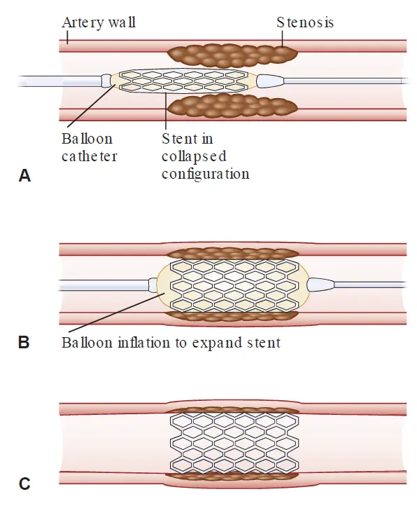
For this reason, coronary stents were developed for implantation during PCI, and have been shown to significantly reduce the rate of restenosis. These stents are cage-like metal devices that in their collapsed state, can be threaded into the region of stenosis by a catheter. Once in position, the stent is expanded into its open configuration by inflating a high-pressure balloon inside it. The balloon and attached catheter are then removed, but the stent is left permanently to maintain coronary patency. Because stents are thrombogenic, oral antiplatelet agents are crucial after stenting.
Although restenosis resulting from vessel elastic recoil is greatly diminished by standard metal stent placement, neointimal proliferation (i.e. migration of smooth muscle cells and production of extracellular matrix) remains an important cause of in-stent restenosis and recurrent anginal symptoms. To address the problem, drug-eluting stents were developed. These stents are manufactured using a polymer coated with an anti-proliferative medication such as sirolimus (immunosuppressive agent that inhibits T-cell activation), everolimus (immunosuppressive similar to sirolimus), or paclitaxel (interferes with cellular microtubule function). The medication is released over a period of 2 to 4 weeks, and this approach has shown great effect at preventing neointimal proliferation and reducing the need for repeat revascularization by more than half.
However, just as neointimal proliferation is slowed, so too is protective endothelialization of the stent. The delay in endothelial cell coverage of the stent leaves patients at risk for in-stent thrombus formation, especially if antiplatelet agents be discontinued prematurely. Thus, prolonged courses of combination antiplatelet therapy followed by indefinite aspirin administration are necessary for patients who receive drug-eluting stents.
Although percutaneous revascularization is generally superior to standard medical therapy for relief of angina, it is important to note that in the setting of stable coronary disease, they have not been shown to reduce the risk of MI or death.
CABG
CABG surgery involves grafting portions of a patient’s native blood vessel to bypass obstructed coronary arteries. One type of grafting used employs native veins—typically, a section of the saphenous vein that is sutured from the base of the aorta to a coronary segment downstream from the region of stenosis. Another method of grafting uses arterial grafts, most commonly, an internal mammary artery (IMA) that can be directly anastomosed distal to a stenotic coronary site.
Vein grafts have a patency rate of up to 80% at 12 months but are vulnerable to accelerated atherosclerosis; 10 years after surgery, more than 50% have occluded. In contrast, IMA grafting is more resistant to atherosclerosis with a patency rate of 90% at 10 years. Therefore, IMA grafts are often used to perfuse sites of critical flow such as the left anterior descending artery.
Clinical trial evidence supports the use of aggressive lipid-lowering drug therapy after CABG to improve the long-term patency rates of bypass grafts.
In recent years, less invasive surgical alternatives to CABG have been explored. These include “minimally invasive” operations with smaller incisions, the use of transcutaneous ports with videoscopic robotic assistance, and “off-pump” procedures, which avoid the use of cardiopulmonary bypass (heart–lung) machines.
While there are theoretical advantages to avoiding CABG, studies examining off-pump procedures in comparison with standard CABG have shown comparable mortality benefit, but poorer graft patency over time and an increased need for future revascularization. Additionally, there have been no major high-quality studies comparing benefits of minimally invasive operations to conventional CABG. In general, patient-specific risks and characteristics are considered by the surgeon when selecting the type of operation to undertake.
See also
References
Additional Reading
- Bonaca MP, Bhatt DL, Cohen M, et al. Long-term use of ticagrelor in patients with prior myocardial infarction. N Engl J Med. 2015;372:1791–1800.
- Douglas PS, Hoffmann U, Patel MR, et al. Outcomes of Anatomical versus Functional Testing for Coronary Artery Disease. N Engl J Med. 2015;372:1291–1300.
- Farkouh ME, Domanski M, Sleeper LA, et al. FREEDOM Trial Investigators. Strategies for multivessel revascularization in patients with diabetes. N Engl J Med. 2012;367:2375–2384.
- Fihn SD, Gardin JM, Abrams J, et al. 2012 ACCF/AHA/ACP/AATS/PCNA/SCAI/STS Guideline for the diagnosis and management of patients with stable ischemic heart disease: executive summary. Circulation. 2012;126:3097–3137.
- Levine GN, Bates ER, Blankenship JC, et al. 2011 ACCF/AHA/SCAI guideline for percutaneous coronary intervention: executive summary. Circulation. 2011;124:2574–2609.
- Mohr FW, Morice MC, Kappetein AP, et al. Coronary artery bypass graft surgery versus percutaneous coronary intervention in patients with three-vessel disease and left main coronary disease: 5-year follow-up of the randomized, clinical SYNTAX trial. Lancet. 2013;381(9867):629–638.
- Park S-J, Ahn J-M, Kim Y-H, et al. Trial of everolimus-eluting stents or bypass surgery for coronary disease. N Engl J Med. 2015;372:1204–1212.
- Tonino PA, De Bruyne B, Pijls NH, et al. FAME Study Investigators. Fractional flow reserve versus angiography for guiding percutaneous coronary intervention. N Engl J Med. 2009;360:213–224.
- Velazquez EJ, Lee KL, Deja MA, et al. STITCH Investigators. Coronary-artery bypass surgery in patients with left ventricular dysfunction. N Engl J Med. 2011;364:1607–1616.