Part 13: Peripheral Vascular Disease
Thu Aug 06 2026
By B. Hassan
Peripheral vascular disease is an umbrella term that includes a number of diverse pathologic entities that affect blood vessels. Although this terminology implies a distinction between the “central” coronary and “peripheral” systemic vessels, the vasculature as a whole comprises a dynamic, integrated system that does not naturally comply with this semantic division.
Peripheral vascular diseases result from 3 categories of pathologies: (1) structural changes in the vessel wall secondary to degenerative conditions, infection, or inflammation that lead to dilatation, aneurysm, dissection, or rupture; (2) narrowing of the vascular lumen caused by atherosclerosis, thrombosis or inflammation; and (3) spasm of vascular smooth muscle. These can occur in isolation or in combination.
The aorta
The aorta is the largest vessel of the vascular system. In adults, its diameter is cm at its origin at the base of the heart. The ascending aorta, 5 to 6 cm in length, leads to the aortic arch, from which arise three major branches: the brachiocephalic artery, the left common carotid artery, and the left subclavian artery. The brachiocephalic artery then bifurcates into the right common carotid and subclavian arteries.
As the descending aorta continues beyond the arch, its diameter narrows to approximately 2 to 2.5 cm in healthy adults. As the aorta pierces the diaphragm, it becomes the abdominal aorta, providing arteries to the abdominal viscera before bifurcating into the left and right common iliac arteries, which supply the pelvic organs and lower extremities.
The aorta is subject to injury from mechanical trauma because it is continuously exposed to high pressure and shear stress. The predominance of elastin in the media allows the aorta to expand during systole and recoil during diastole. This recoil against the closed aortic valve contributes to the distal propagation of blood during ventricular relaxation. With advancing age, the elastic component of the aorta and its branches degenerates and collagen becomes more prominent, causing arterial stiffening. Systolic blood pressure (SBP), therefore, tends to rise with age because less energy is dissipated into the aorta during left ventricular contraction.
Aortic aneurysm
An aneurysm is an abnormal dilatation of an artery. In the aorta, aneurysms are distinguished from diffuse ectasia, which is a generalized yet lesser increase of the aortic diameter.
Ectasia develops in older patients as elastic fibers and smooth muscle cells decrease in number, while mucopolysaccharide ground substances accumulate within the vessel wall.
The term aneurysm is applied when the diameter of a portion of the aorta has increased by at least 50% compared with normal. A true aneurysm represents a dilatation of all three layers of the aorta and is characterized as either fusiform or saccular, depending on the extent of the vessel’s circumference within the aneurysm. A fusiform true aneurysm (more common) is characterized by symmetrical dilation of the entire circumference of a segment of the aorta. A saccular true aneurysm is a localized outpouching involving only a portion of the circumference.
In contrast, a pseudoaneurysm (or false aneurysm) is a contained breach of all layers of the arterial wall. Blood leaks out of the vascular lumen through a defect in the intima, media, and adventitia, and is contained only by adventitial tissue remnants, perivascular connective tissue, or a thrombus. Pseudoaneurysms develop at sites of vessel injury caused by infection or trauma, such as puncture of the vessel during surgery or percutaneous catheterization. They are very unstable and are prone to rupture.
Aneurysms may be confined to the abdominal aorta (most common), the thoracic aorta, or involve both locations. They may also appear in peripheral and cerebral arteries.
Etiology and Pathogenesis of True Aortic Aneurysms
The etiology of aortic aneurysm varies depending on the location. Ascending thoracic aortic aneurysms are typically characterized by cystic medial degeneration, a condition of degeneration of elastic fibers with subsequent accumulation of collagenous and mucoid material within the medial layer.
Cystic medial degeneration occurs normally with age but is also associated with hypertension. Additionally, it develops in certain inherited connective tissue disorders including Marfan syndrome, Loeys–Dietz syndrome, the vascular form of Ehlers–Danlos syndrome (type IV), and familial thoracic aortic aneurysms. Cystic medial degeneration also characterizes the form of thoracic aortic aneurysm often associated with bicuspid aortic valves.
Aneurysms of the descending thoracic and abdominal aorta are usually associated with atherosclerosis and its risk factors, including smoking, hypertension, dyslipidemia, male gender, and advanced age. However, it is unlikely that atherosclerosis alone is responsible for such aneurysm. Rather, other pathophysiologic mechanisms including genetic predisposition, local vessel inflammation, and an imbalance between synthesis and degradation of extracellular matrix proteins are also implicated.
Aneurysms are also associated with markers of inflammation, including CRP and cytokines such as IL-6, which have been shown to correlate with the size of aneurysms. Moreover, inflammatory cells such as lymphocytes and macrophages are frequently found on histologic examination. Angiotensin II, via its effect on inflammation and oxidative stress, has also been implicated in experimental models of abdominal aortic aneurysms.
Infrequent causes of aortic aneurysms include weakness of the media due to infections of the vessel wall and vasculitis.
Clinical Presentation and Diagnosis
Most aneurysms are asymptomatic, though some patients, especially those with abdominal aortic aneurysms, may present with a pulsatile mass. Others present with symptoms related to compression of neighboring structures. For example, thoracic aortic aneurysms may compress the trachea or mainstem bronchus, causing cough or dyspnea. Compression of the esophagus can result in dysphagia, and involvement of the recurrent laryngeal nerve may lead to hoarseness. Aneurysms of the ascending aorta may dilate the aortic ring, resulting in aortic regurgitation. Abdominal aortic aneurysms may cause abdominal or back pain or non-specific gastrointestinal symptoms.
Aortic aneurysms are often first suspected when dilatation of the vessel is observed on radiography. Aneurysms of the abdominal aorta or of large peripheral arteries may also be discovered by careful palpation during physical examination. The diagnosis is confirmed by ultrasonography, contrast-enhanced CT, or MRI.
The most devastating consequence of an aortic aneurysm is rupture, which can be fatal. An aneurysm may leak slowly or burst suddenly, resulting in profound bleeding and hypotension. Thoracic aortic aneurysms may rupture into the pleural space, mediastinum, or bronchi. Abdominal aortic aneurysms may rupture into the retroperitoneal space or abdominal cavity or erode into the intestines, resulting in massive gastrointestinal bleeding.
The following CT angiography shows an abdominal aortic aneurysm:
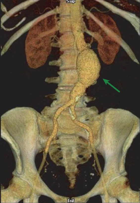
Studies have shown that the risk of rupture is related to the size of the aneurysm, as predicted by the Laplace relationship (i.e. wall tension is proportional to the pressure and the radius). The mean rates of thoracic and abdominal aortic aneurysms expansion are 0.1 and 0.4 cm/ year, respectively.
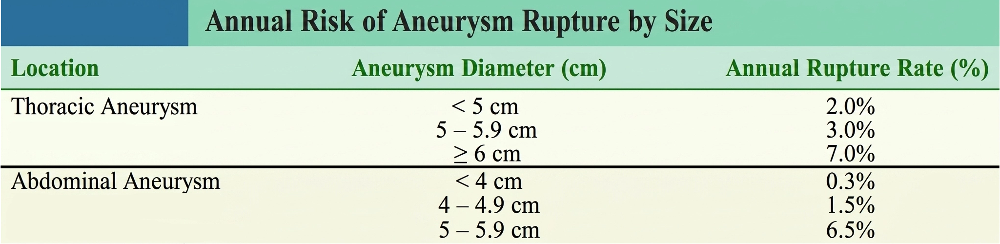
The annual rupture rates for aneurysms of different diameters are described in the following table:
Note
Abdominal aneurysms cm have a markedly higher risk of rupture
Treatment
Treatment of an aortic aneurysm is based on its size and the patient’s overall condition. Once an aneurysm is identified, its size should be closely monitored through repeated imaging, typically every 6 to 12 months depending on size. In general, surgical treatment is considered for ascending aortic aneurysms greater than 5.5-6.0 cm. Ascending aortic aneurysms in patients with Marfan syndrome should be considered for surgical repair if the diameter is cm. Surgical repair is generally recommended for descending thoracic aortic aneurysms measuring 6.5-7.0 cm, or abdominal aortic aneurysms measuring cm, or smaller aneurysms that enlarge at a rate cm/year.
The mortality associated with elective surgical repair of thoracic aortic aneurysms is 3% to 5%. Patients are maintained on cardiopulmonary bypass as the aneurysm is resected and replaced with a prosthetic Dacron graft. Patients with aneurysms involving multiple aortic segments have staged repairs, in which one segment is corrected at a time. Some patients with thoracic aortic aneurysms may be candidates for minimally invasive repair, in which an endovascular stent graft is positioned across the aneurysm.
Surgical repair of abdominal aortic aneurysms involves placement of a prosthetic graft. The operative mortality for such procedures at high-volume institutions is 1% to 2%. Percutaneous endovascular repair of infrarenal abdominal aortic aneurysms with stent grafts can be performed in selected patients with less acute morbidity, and long-term results are similar to that of surgical repair.
Medical management, including risk-factor reduction, is currently recommended for patients with small aneurysms. β-Blockers may reduce the expansion rate of thoracic aortic aneurysms in patients with Marfan syndrome; it is not clear whether they are effective for other causes or types of aneurysms.
Aortic dissection
Aortic dissection arises from a tear in the intimal layer of the vessel wall that allows blood from the lumen to enter into the media and propagate along the plane of the muscle layer. Another potential origin of aortic dissection is rupture of vasa vasorum with hemorrhage into the media, forming a hematoma in the arterial wall that subsequently tears through the intima and into the vessel’s lumen. Other related acute syndromes include aortic intramural hematoma, penetrating aortic ulcer, and aortic rupture.
Acute intramural hematoma is a variant of aortic dissection characterized by a hemorrhage in the wall of the aorta without evidence of an intimal tear. A penetrating atherosclerotic ulcer results from erosion of a plaque into the aortic wall. Aortic rupture may be a complication of aortic dissection, intramural hematoma, penetrating atherosclerotic ulcer, or from trauma.
Any condition that interferes with the normal integrity of the elastic or muscular components of the medial layer can predispose to aortic dissection. Such degeneration may arise from chronic hypertension, aging, and/ or cystic medial degeneration. In addition, traumatic damage to the aorta (e.g. blunt chest trauma or accidental damage during intra-arterial catheterization or cardiac surgery) can also initiate dissection.
Aortic dissection is most common in the sixth and seventh decades and occurs more frequently in men. More than two thirds of patients have a history of hypertension.
Dissections are commonly classified into two categories (Stanford type A and Stanford type B), depending on their location and extent. In a type A dissection (proximal), the ascending aorta and/ or aortic arch is involved, regardless of the site of the primary tear. Type B dissection (distal) does not involve the ascending aorta or arch and is, therefore, confined to the descending thoracic and abdominal aorta. This distinction is important because treatment strategies and prognosis are determined by location. Proximal aortic involvement tends to be more devastating because of the potential for extension into the coronary and arch vessels, the support structures of the aortic valve, or the pericardial space. Approximately two thirds of dissections are type A and one third are type B, with primary isolated abdominal aortic dissections being extremely rare (<1–2%).
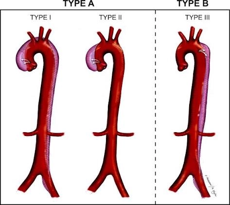
Dissections may also be classified as acute or chronic, with acute dissections presenting with symptoms of less than 2 weeks’ duration.
Clinical Presentation and Diagnosis
The most common symptom of aortic dissection is sudden severe “tearing” pain in the anterior chest (typical of type A dissections) or between the scapulae (type B dissections). The pain travels as the dissection propagates along the aorta and can radiate anywhere in the thorax or abdomen. Painless dissection is possible but uncommon (generally of cases).
Other symptoms relate to the complications that can occur with the tear. These include rupture anywhere along the aorta (often into the pleural space or pericardium); occlusion of major branches of the aorta by the hematoma within the vessel wall, which can result in myocardial infarction (coronary artery involvement), stroke (carotid artery involvement), visceral ischemia, renal failure, or loss of peripheral pulsation in an extremity; and extension into the aortic root, disrupting the aortic valve support apparatus causing aortic regurgitation.
Several important physical findings may be present. Hypertension is frequently detected, either as an underlying cause of dissection, a result of the sympathetic nervous system response to the severe pain, or because of diminished renal flow with activation of the renin–angiotensin system. If the dissection has occluded one of the subclavian arteries, a difference in SBP between the arms is noted. Neurologic deficits may accompany dissection into the carotid vessels. If a type A dissection results in aortic regurgitation, an early diastolic murmur can be detected on auscultation. Leakage from a type A dissection into the pericardial sac may produce signs of cardiac tamponade.
The diagnosis of aortic dissection must not be delayed, because catastrophic complications may rapidly ensue. The confirmatory imaging techniques most useful in detecting dissection include contrast-enhanced CT, transesophageal echocardiography, and contrast angiography. Each of these techniques has specific advantages and disadvantages, and the decision of which to employ is often guided by a hospital’s local expertise.
Treatment
The goal of acute treatment is to arrest progression of the dissecting blood stream. Suspicion of acute aortic dissection warrants immediate medical therapy to reduce SBP (aiming for a SBP of 100 to 120 mmHg) and to decrease left ventricular contraction and thus minimize aortic wall shear stress. Useful pharmacologic agents to achieve this include β-blockers and vasodilators.
In type A dissections, early surgical correction has been shown to improve outcomes compared with medical therapy alone. Surgical therapy involves repairing the intimal tear, suturing the edges of the false lumen, and if necessary, inserting a synthetic aortic graft.
In contrast, patients with uncomplicated type B dissections are initially managed with aggressive medical therapy alone; early surgical intervention does not further improve the outcome in these patients. Surgery is indicated, however, if there is clinical evidence of propagation of the dissection, compromise of major branches of the aorta, impending rupture, or continued pain. Percutaneous catheter-based repair with endovascular stent grafts has been used successfully in selected stable patients with type B dissections. The graft seals the entry site of the dissection, resulting in thrombosis of the false lumen.
Peripheral Atherosclerotic Vascular Disease
The most common cause of peripheral artery disease (PAD) is atherosclerosis. It affects approximately 4% of people over 40 and 15% to 20% of those over 70. The pathology of atherosclerotic PAD is identical to that of coronary artery disease (CAD), and the major risk factors are also the same. Approximately 40% of patients with PAD actually have clinically significant CAD. Thus, detection of PAD is useful in identifying patients at increased risk of adverse cardiovascular events.
Ischemia of the affected region occurs when the balance between oxygen supply and demand is upset; exercise raises the demand for blood in the limbs’ muscles, and a stenosed or obstructed artery cannot provide an adequate supply. Rest improves symptoms as the balance between oxygen supply and demand is restored.
Recall from part 5 that the degree of blood flow reduction relates to the extent of vessel narrowing, the length of the stenosis, and blood viscosity according to the Poiseuille equation, and that the degree of vessel narrowing by the stenosis (i.e. the change in radius) has the greatest impact on flow. The equation also indicates that as the flow velocity increases across a stenotic vessel, the blood turbulence results in a loss of kinetic energy. The result is a decline in perfusion pressure distal to the stenosis.
During exercise, metabolites (e.g. adenosine) act locally to dilate arterioles, decreasing vascular resistance, which serves to increase blood flow to the active muscle. In turn, the increased flow stimulates healthy arterial endothelium to release vasodilating factors such as nitric oxide, thereby increasing the radii of upstream vessels. However, in PAD, obstructed arteries cannot respond to the vasodilating stimuli, thereby limiting flow increases. In addition, dysfunctional atherosclerotic endothelium does not release normal amounts of vasodilating substances. Thus, the physical properties of a stenosis and the reduced vasodilator activity imposed by diseased endothelium prevent adequate blood flow from reaching distal tissues and contribute to ischemia.
Hemodynamic changes alone cannot account for the dramatic reductions in exercise capacity experienced by PAD patients; changes in muscle structure and function are also seen. One such change is the denervation of muscle fibers, resulting in muscle atrophy, which is thought to occur as an adaptation to intermittent ischemia. Even viable muscle fibers in an affected limb may show abnormalities of mitochondrial oxidative metabolism.
Clinical Presentation and Diagnosis
PAD may affect the iliac, femoral, popliteal, and tibioperoneal arteries. Patients with PAD may therefore develop buttock, thigh, or calf discomfort precipitated by walking and relieved by rest. This classic symptom of exertional limb fatigue and pain is known as claudication. In severe PAD, patients may experience pain at rest.
Note
The femoral and popliteal arteries being the most common sites. The arteries of the upper extremities are less frequently affected, but brachiocephalic or subclavian artery disease can cause arm claudication.
Physical examination generally reveals loss of pulse distal to the stenosis. Bruits may also be heard in the affected region. In patients with chronic severe ischemia, the lack of blood perfusion results in muscle atrophy, pallor, cyanotic discoloration, hair loss, and occasionally gangrene and necrosis of the foot and digits.
Ischemic ulcers resulting from PAD often begin as small traumatic wounds in areas of pressure or in regions prone to injury, such as the tips of the toes and the volar surface of the foot. These often painful ulcers fail to heal owing to the inadequate blood supply. Diabetic patients with peripheral sensory neuropathies are particularly susceptible to ulcers at sites of trauma or pressure from ill-fitting footwear. Ischemic ulcers can be distinguished from venous insufficiency ulcers, which develop more proximally and on the medial portion of the leg. Venous ulcers are also associated with reddish-brown pigmentation and varicose veins.
In the evaluation of PAD, it is helpful to measure the ratio of blood pressure in the ankles to that in the arms (termed the ankle–brachial index or ABI). Other testing to assess peripheral perfusion includes limb segmental systolic pressure measurements and pulse volume recordings (i.e. graphical measurement of volume changes in segments of the extremity with each pulse). Duplex ultrasonography is a commonly used noninvasive method to visualize and assess the extent of arterial stenoses and the corresponding reductions in blood flow. Other more advanced imaging studies (e.g. MRI angiography, CT angiography, or intra-arterial contrast angiography) are obtained when revascularization procedures are planned.
Treatment
For patients with PAD, antiplatelet therapy and risk factor modification (smoking cessation, lipid lowering, and control of diabetes and hypertension) are important in mitigating coronary events. Antiplatelet agents such as aspirin and clopidogrel has been shown to reduce cardiovascular morbidity and mortality in patients with PAD, though it has not been established if antiplatelet agents reduce symptoms or prevent thrombotic complications of PAD itself.
Specific treatment of PAD includes appropriate foot care to prevent trauma or restriction of blood flow and exercise (particularly walking) which increases metabolic efficiency in the skeletal muscle of the legs. A formal exercise program is considered first-line therapy in the management of PAD.
Certain medications are sometimes useful in the treatment of claudication. Cilostazol is a selective phosphodiesterase inhibitor that increases cAMP and has vasodilator and platelet-inhibiting properties; it has been shown to improve exercise capacity in patients with PAD. Pentoxifylline is a drug purported to improve the deformability of red and white blood cells and may improve claudication symptoms in some patients. Conversely, most vasodilator drugs are not helpful in relieving claudication.
More effective medical therapies for PAD are on the horizon. Advances in angiogenesis research and clinical trials provide hope that revascularization through delivery of angiogenic growth factors and regenerative cell-based therapies, including use of endothelial progenitor cells, may be possible.
Revascularization is indicated when medical therapy has failed or in patients with disabling claudication and as first-line therapy in cases of critical limb ischemia. Catheter-based interventions, such as percutaneous transluminal angioplasty and stent implantation, can be performed on selected patients with low morbidity. Surgical procedures include bypass operations to circumvent the occluded arteries using saphenous vein or prosthetic grafts. However, amputation may be necessary if blood flow cannot be satisfactorily reestablished to maintain limb viability.
Acute Arterial Occlusion
Acute arterial occlusion is caused either by embolization from a distant site or by thrombosis in situ. The origin of arterial emboli is most often the heart, usually resulting from disorders involving intracardiac stasis of blood. Primary arterial thrombosis may appear at sites of endothelial damage or atherosclerotic stenoses, or within bypass grafts.
Rarely, arterial emboli originate from the venous circulation. If a venous clot travels to the right-heart chambers and is able to pass through an abnormal intracardiac communication (e.g. an atrial septal defect), it then enters the systemic arterial circulation (a condition known as paradoxical embolism).
The extent of tissue damage from thromboembolism depends on the site of the occluded artery, the duration of occlusion, and the degree of collateral circulation serving the tissue beyond the obstruction. Common symptoms and signs that may develop from abrupt occlusion include Pain, Pallor, Paralysis, Paresthesia, and Pulselessness (termed the “five Ps”). A sixth P, Poikilothermia (coolness), is also often present.
Patients with a proven acute arterial occlusion should be treated with a parenteral anticoagulant such as heparin (followed by oral warfarin) to prevent propagation of the clot and to reduce the risk of additional embolic events. A revascularization procedure (catheter-based thrombolysis or thrombectomy, surgical embolectomy, or bypass surgery) is indicated if limb viability is at risk.
Atheroembolism is the condition of peripheral small arterial occlusion by atheromatous material (i.e. cholesterol, platelets, and fibrin) derived from more proximal vascular atherosclerotic sites. Patients typically present with acute pain and tenderness at the involved site. Occlusion of digital vessels may result in the “blue toe” syndrome, culminating in gangrene and necrosis. Other findings may include livedo reticularis (purplish mottling of involved skin), kidney failure (renal atheroembolism), and intestinal ischemia. Although an estimated 50% to 60% of cases are spontaneous, atheroembolism may occur after intravascular procedures (e.g. cardiac catheterization) when atherosclerotic material is unintentionally dislodged. Ischemia resulting from atheroemboli is difficult to manage because the heterogeneous composition and distribution of emboli often precludes surgical removal or thrombolytic therapy. Surgical intervention to remove or bypass the source of emboli may be necessary to prevent recurrences.
Vasculitis
Vasculitis (vessel wall inflammation) results from immune complex deposition or cell-mediated immune reaction against the vessel wall. Immune complexes activate the complement system with subsequent release of chemoattractant agents and anaphylatoxins that direct neutrophils to the vessel wall and increase vascular permeability. Neutrophils injure the vessel by releasing lysosomal contents and producing oxygen-derived free radicals.
In cell-mediated immune reactions, T lymphocytes bind to vascular antigens and release lymphokines that attract additional lymphocytes and macrophages. These inflammatory processes can cause end-organ ischemia through vascular necrosis or local thrombosis.
The cause of most of the vasculitic syndromes is unknown, but they often can be distinguished from one another by the pattern of involved vessels and by histologic characteristics. Three important examples of vasculitis are Takayasu arteritis, giant cell arteritis, and thromboangiitis obliterans.
Takayasu arteritis
Takayasu arteritis is a chronic vasculitis of unknown etiology that targets the aorta and its major branches.
Between 80% and 90% of affected individuals are women, with onset typically between the ages 10 and 40. Most reported cases have been from Asia and Africa, but it is a worldwide disease.
Patients typically present with constitutional symptoms such as malaise and fever. Local symptoms depend on the affected vessel and include cerebrovascular ischemia (brachiocephalic or carotid artery involvement), myocardial ischemia (coronary artery), arm claudication (brachiocephalic or subclavian artery), or hypertension (renal artery).
The carotid and limb pulses are diminished or absent in nearly 85% of patients with Takayasu arteritis at the time of diagnosis; hence it is often termed “pulseless” disease. Takayasu arteritis is also an uncommon cause of aortic aneurysm or aortic dissection.
Histologic examination of affected vessels reveals granulomatous inflammation resulting in intimal proliferation, disruption of the elastic lamina, and fibrosis. Anti-endothelial antibodies may also play a role in the disease.
Steroid and cytotoxic drugs may reduce vascular inflammation and alleviate the symptoms. Surgical bypass of obstructed vessels may be helpful in severe cases. The 5-year survival rate is 80% to 90%.
Giant cell arteritis
Giant cell arteritis (also termed temporal arteritis) is a chronic vasculitis of medium-sized to large arteries that most commonly involves the cranial vessels or the aortic arch and its branches.
It is an uncommon disease and the typical age of onset is after 50, with 65% of patients are female. It may be associated with polymyalgia rheumatica.
Histologic findings in affected vessels include lymphocyte and macrophage infiltration, intimal fibrosis, and focal necrosis, with granulomas containing multinucleated giant cells.
Symptoms and signs depend on the affected arteries and may include diminished temporal pulses, prominent headache, facial pain, and jaw claudication while chewing (facial artery involvement). Ophthalmic artery involvement leads to impaired vision.
Serum markers of inflammation (e.g. ESR and CRP) are invariably elevated. Ultrasonography can support the diagnosis by demonstrating a hypoechoic halo around the involved arterial lumen with vessel stenosis. The diagnosis can be confirmed by biopsy of an involved vessel, usually a temporal artery, but treatment should not wait for biopsy results.
High-dose steroids are effective in treating vasculitis and preventing visual complications. Giant cell arteritis usually has a self-limited course of 1 to 5 years.
Thromboangiitis obliterans
Thromboangiitis obliterans (also called Buerger disease) is a segmental inflammation of small and medium-sized arteries, veins, and nerves involving the distal vessels of the extremities.
It is most prevalent in the Far and Middle East and has a very strong association with cigarette smoking. It is most common in men younger than age 45. There is an increased incidence of HLA-A9 and HLA-B5 in affected individuals.
Thromboangiitis obliterans presents with distal arterial occlusion, Raynaud phenomenon, and migrating thrombophlebitis. Arterial occlusion results in limb claudication as well as ischemia of the digits.
Traditional laboratory markers of inflammation and autoimmune disease are usually not detected. Features of involved arteries include areas of stenosis interspersed with normal-appearing vessels with more severe disease distally and lack of atherosclerosis in proximal arteries.
The diagnosis can be established by tissue biopsy, although this is rarely needed. Biopsy reveals an occlusive, highly cellular, inflammatory thrombus, with limited involvement of the vessel wall.
The most important treatment in thromboangiitis obliterans is smoking cessation, which usually prevents progression of the disease and its complications. Debridement of necrotic tissue may be necessary in advanced cases. Revascularization is not usually an option because of the distal location of the arterial lesions.
Venous disease
Veins are high-capacitance vessels that contain more than 70% of the total blood volume. In contrast to the muscular structure of arteries, the subendothelial layer of veins is thin, and the tunica media comprises fewer bundles of smooth muscle cells intermixed with reticular and elastic fibers.
While veins of the extremities possess intrinsic vasomotor activity, transport of blood back to the heart relies greatly on external compression by the surrounding skeletal muscles and on a series of one-way valves.
Veins of the extremities are classified as either deep or superficial. In the lower extremities, where most peripheral venous disorders occur, the deep veins generally course along the arteries, whereas the superficial veins are located subcutaneously. The superficial veins drain into deeper veins through a series of perforating connectors.
Varicose Veins
Varicose veins are dilated tortuous superficial veins that often develop in the lower extremities. Clinically apparent varicose veins occur in 10% to 20% of the general population. They affect women 2-3 times more frequently than men, and roughly half of patients have a positive family history. Varicosities can occur in any vein in the body but are most common in the saphenous veins of the leg and its tributaries. They may also develop in the anorectal area (hemorrhoids), in the lower esophageal veins (esophageal varices), and in the spermatic cord (varicocele).
Varicosity is thought to result from intrinsic weakness of the vessel wall, from increased intraluminal pressure, or from congenital defects in venous valves. Varicose veins in the lower extremities are classified as either primary or secondary.
Primary varicose veins originate in the superficial system, and factors that lead to their development include pregnancy, prolonged standing, and obesity. During pregnancy or prolonged standing, the high venous pressure within the legs contributes to varicosities when there is underlying weakness of the vessel walls. In obese patients, the adipose tissue surrounding vessel offers less structural support to veins than does lean mass.
Secondary varicose veins occur when an abnormality in the deep venous system is the cause of superficial varicosities. These may develop in the setting of deep venous insufficiency, occlusion, or when the perforating veins are incompetent. In such cases, deep venous blood is shunted retrogradely through perforating channels into superficial veins, increasing intraluminal pressure and volume and causing dilatation and varicosity formation.
Many people with varicose veins are asymptomatic but seek treatment for cosmetic reasons. When symptoms do develop, they include a dull ache pressure sensation in the legs after prolonged standing. Superficial venous insufficiency may result when venous valves are unable to function normally in the dilated veins. This can cause swelling and skin ulceration. Stasis of blood within varicose veins can promote superficial vein thrombosis, and varicosities can also rupture, causing a localized hematoma.
Varicose veins are usually treated conservatively. Patients should elevate their legs while supine, avoid prolonged standing, and wear external compression stockings that counterbalance the increased venous hydrostatic pressure. Varicose veins that are symptomatic may be treated with more advanced techniques, including sclerotherapy, thermal ablation, or surgery. Sclerotherapy involves the localized intravenous administration of an irritating agent to fibrose varicose veins. Endovenous thermal ablation uses laser or radio frequency catheters to deliver heat energy, which induces thromboses and is used to obliterate varicose great saphenous veins.
Deep Venous Thrombosis
Deep venous thrombosis (DVT) occurs most commonly in the veins of the calves but may also develop initially in more proximal veins such as the popliteal, femoral, and iliac vessels. If left untreated, 20% to 30% of DVTs that arise in the calves propagate to these proximal veins. The two major consequences of DVT are pulmonary embolism (PE) (also termed venous thromboembolism) and postphlebitic syndrome.
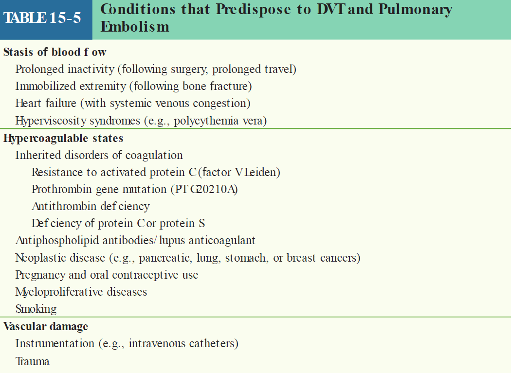
The classic triad of factors that predispose to venous thrombosis is stasis of blood flow, hypercoagulability, and vascular damage. Stasis disrupts laminar flow and brings platelets into contact with the endothelium, allowing coagulation factors to accumulate. Factors that slow venous flow and induce stasis include prolonged immobilization (e.g. bed rest after surgery or sitting in a car or an airplane for a long trip), cardiac failure, and hyperviscosity syndromes.
Various clinical disorders cause systemic hypercoagulability, including resistance of coagulation factor V to activated protein C, a prothrombin gene mutation, and inherited deficiencies of antithrombin, protein C, and protein S. Pancreatic, lung, stomach, breast, and genitourinary tract adenocarcinomas are associated with a high prevalence of venous thrombosis. This is thought to occur in part because necrotic tumor cells release thrombogenic factors.
Vascular damage, either by external injury or by IV catheters, can expose subendothelial collagen, which in turn acts as a substrate for the binding of von Willebrand factor and platelets to initiate the clotting cascade. Less severe damage can cause endothelial dysfunction that contributes to thrombosis by disrupting the production of natural vasodilating and antiplatelet substances. Recent evidence indicates that atherosclerotic risk factors, such as hyperlipidemia and diabetes, are also associated with DVT formation.
The risk of DVT is particularly high after fractures of the spine, pelvis, and bones of the lower extremities, which may be related to stasis of blood flow, increased coagulability, and possibly traumatic endothelial damage. In addition, venous thrombosis may arise in patients following surgical procedures, particularly major orthopedic operations.
Women have a several-fold increase in the incidence of DVT during late pregnancy and the early postpartum period. In the third trimester, the fetus compresses the inferior vena cava and can cause stasis of blood flow, and high levels of circulating estrogen may induce a hypercoagulable state. Oral contraceptives and other pharmacologic estrogen products also predispose to thrombus formation.
Clinical Presentation and Diagnosis
Patients with DVT may be asymptomatic, may describe calf or thigh discomfort when standing or walking, or may report unilateral leg swelling. The physical findings of proximal DVT include edema of the involved leg and occasionally localized warmth and erythema. Tenderness may be present over the course of the vein, and a deep venous cord is occasionally palpable. Calf pain produced by dorsiflexion of the foot (the Homan sign) is a non-specific and unreliable marker of DVT.
The primary laboratory tests for the diagnosis of DVT include measurement of the serum d-dimer level and venous compression ultrasonography.
d-dimer is a by-product of fibrin degradation that can be measured in a peripheral blood sample and is highly sensitive for the diagnosis of DVT and/or acute PE. Because d-dimer may also be elevated in many other conditions (such as cancer, inflammation, infection, and necrosis), a positive test result is not specific for DVT. Thus, a normal d -dimer value helps exclude the presence of DVT, but an elevated level does not definitively confirm the diagnosis.
Venous compression duplex ultrasonography is a readily available noninvasive technique that is 95% sensitive for the diagnosis of symptomatic DVT in a proximal vein but only 75% sensitive for diagnosing symptomatic calf vein thrombi. Criteria used for diagnosis of DVT with duplex ultrasonography include the inability to compress the vein with direct pressure (suggesting the presence of an intraluminal thrombus), direct visualization of the thrombus, and absence of blood flow within the vessel.
Other diagnostic techniques are sometimes used. For example, MRI venography can aid in the diagnosis of proximal DVT, particularly pelvic vein thrombi, which are difficult to detect by ultrasound. Contrast venography is now a rarely used invasive imaging technique that can provide a definitive diagnosis. Radiocontrast material is injected into a foot vein, and images are obtained as the contrast ascends through the venous system of the leg. DVT is diagnosed by the presence of a filling defect.
Treatment
In patients with proximal DVT, elevation of the affected extremity above the level of the heart helps reduce edema and tenderness, and anticoagulation prevents extension of the thrombus and PE. Initial anticoagulation typically consists of low molecular weight heparin (LMWH). IV unfractionated heparin is a cost-effective alternative that has been used successfully for this purpose for many years, but LMWH is more convenient to administer.
Warfarin, an oral anticoagulant, is then prescribed for long-term management and is continued for several months, depending on the underlying cause of DVT. Newer oral anticoagulants, such as the factor Xa inhibitors rivaroxaban and apixaban allow a broader range of options for acute and long-term management of DVT. Catheter-based thrombolysis may be useful for selected patients with iliofemoral deep vein thrombosis.
Treatment of patients with calf DVT is more controversial because pulmonary emboli from that site are uncommon. Some experts advocate serial non-invasive monitoring to determine if the thrombus propagates into proximal veins, whereas others treat such thrombosis with heparin followed by warfarin for 3 to 6 months.
Prophylaxis against DVT is appropriate in situations in which the risk of developing DVT is high, such as during bed rest following surgery. Prophylactic measures may include unfractionated heparin, LMWH, or one of the newer oral anticoagulants, as well as compression stockings, and/or intermittent external pneumatic compression of the legs to prevent venous stasis.
Pulmonary Embolism
PE occurs when a clot, most often derived from DVT in a proximal vein dislodges and travels through the inferior vena cava and right heart chambers, finally reaching and obstructing a portion of the pulmonary circulation. PE is common and is often fatal, with an untreated mortality rate of 30% to 40%.
When a pulmonary embolism occurs, the blood is shunted into the healthy portions of the lungs, and so the lung vasculature is divided into 2 zones:
-
Zone A (Blocked): This is the portion blocked by the emboli, and so air enters, but no blood. This is Dead Space ventilation.
-
Zone B (Healthy): now receives its own blood PLUS the extra blood that couldn’t go through Zone A causing pulmonary hypertension.
This leads to an increase in pulmonary artery pressure (due to the obstruction in Zone A and the volume overload in Zone B), which in turn means the right ventricle has to push against higher pressure. This may lead to elevation of right ventricular (RV) wall stress (thus oxygen demand), dilatation, and contractile failure, leading to a RV ischemia. Recurrent and chronic PE can cause remodeling of the pulmonary vasculature with pulmonary hypertension leading to right-sided heart failure (termed cor pulmonale).
There is also a V/Q mismatch, in Zone A, there is too much ventilation and little blood (high V/Q ratio) while there is too much blood and not enough ventilation in Zone B (low V/Q). This mismatch predisposes to hypoxemia despite any compensatory attempts at hyperventilation.
Note
Since CO2 removal is more directly dependent on ventilation (diffusion) than oxygen, the hyperventilation leads to respiratory alkalosis.
Clinical Presentation and Diagnosis
Patients with PE may experience dyspnea, pleuritic chest pain (due to pleural irritation), hemoptysis, cough, or syncope (due to reduced cardiac output). Signs may include tachypnea, bronchospasm, and evidence of elevated pulmonary artery pressure, including an accentuated pulmonic component of S2 and jugular venous distention.
Many of the diagnostic tests used for DVT are also useful for diagnosis PE. Additional tests are useful in the evaluation of suspected PE. The most common electrocardiographic abnormality is sinus tachycardia.
There may also be evidence of RV strain (e.g. inverted T waves in leads V1–V4 or an “S1–Q3–T3” pattern: a prominent S wave in lead I, Q wave in lead III, inverted T wave in lead III). RV strain may also produce elevated serum levels of cardiac troponins or B-type natriuretic peptide.
Arterial blood gas analysis may show alkalosis with decreased arterial oxygenation but is insensitive to the diagnosis of PE.
The preferred test to confirm the diagnosis is CT angiography. For patients who cannot tolerate CT angiography, such as those with renal insufficiency or hypersensitivity to contrast agents, radionuclide ventilation–perfusion (V/Q) lung scanning may be obtained instead but is less precise for the diagnosis.
Treatment
In patients with established PE, urgent anticoagulation is instituted to prevent recurrent embolism. Anticoagulation measures are similar to those used in DVT. In patients with proximal DVT or established PE who cannot be treated with anticoagulants (e.g., because of a bleeding disorder), an intravascular filter can be inserted into the inferior vena cava to prevent emboli from reaching the lungs. Occasionally, systemic thrombolytic therapy or surgical pulmonary embolectomy is undertaken for patients with massive PE.
See also
References
Additional Reading
- Braverman AC. Acute aortic dissection: clinician update. Circulation. 2010;122:184–188
- Creager MA, Beckman JA, Loscalzo J, eds. Vascular Medicine: A Companion to Braunwald’s Heart Disease. Philadelphia, PA: Elsevier Saunders; 2013.
- Creager MA, Loscalzo J. Diseases of the aorta. In: Kasper D, Fauci A, Hauser S, et al., eds. Harrison’s Principles of Internal Medicine. 19th ed. New York, NY: McGraw-Hill; 2015, pp.1637–1643
- Creager MA, Loscalzo J. Arterial diseases of the extremities. In: Kasper D, Fauci A, Hauser S, et al., eds. Harrison’s Principles of Internal Medicine. 19th ed. New York, NY: McGraw-Hill; 2015, pp.1643–1650.
- Gloviczki P, Comerota AJ, Dalsing MC, et al. The care of patients with varicose veins and associated chronic venous diseases: clinical practice guidelines of the Society for Vascular Surgery and the American Venous Forum. J Vasc Surg. 2011;53:2S–48S.
- Goldhaber SZ, Bounzameaux H. Pulmonary embolism and deep vein thrombosis. Lancet. 2012;379:1835–1846.
- Gornik HL, Creager MA. Aortitis. Circulation. 2008;117:3039–3051.
- Hiratzka LF, et al. ACCF/ AHA 2010 guidelines for the diagnosis and management of patients with thoracic aortic disease. J Am Coll Cardiol. 2010;55:e27–e129.
- Patel MR, Conte MS, Cutlip DE, et al. State-of-the-art review: Evaluation and treatment of patients with lower extremity peripheral artery disease. J Am Coll Cardiol. 2015;65:931–941
- Verma S, Siu SC. Aortic dilatation in patients with bicuspid aortic valve. N Engl J Med 2014;370:1920–1929